
Medicine and Healthnpj Regenerative Medicine
Multi-biofunctional graphene oxide-enhanced poly-L-lactic acid composite nanofiber scaffolds for ovarian function recovery of transplanted-tissue
L. Yan, L. Wang, et al.
This research by Liang Yan and colleagues explores innovative graphene oxide/poly-L-lactic acid nanofiber scaffolds that enhance ovarian function in mice with primary ovarian insufficiency. This study not only advances tissue engineering but also presents a fresh approach to organ transplantation and cryopreservation.
Related Publications
Explore these studies to deepen your understanding
Adjacent work that informs or extends this paper's methodology and findings.

Engineering and Technology
Bacterial cellulose-graphene oxide composite membranes with enhanced fouling resistance for bio-effluents management
I. S. Mir, A. Riaz, et al.
Medicine and Health
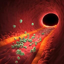
EGFP-EGF1-conjugated poly(lactic-co-glycolic acid) nanoparticles as a carrier for the delivery of CCR2– shRNA to atherosclerotic macrophage in vitro
Z. Wu, C. Chen, et al.

Food Science and Technology
Compact analytical flow system for the simultaneous determination of L-lactic and L-malic in red wines
P. Giménez-gómez, M. Gutiérrez-capitán, et al.

Medicine and Health
Manufacturing micropatterned collagen scaffolds with chemical-crosslinking for development of biomimetic tissue-engineered oral mucosa
A. Suzuki, Y. Kodama, et al.


