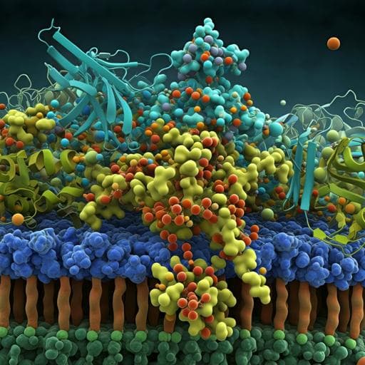
logo
ResearchBunny Logo
Introduction
Membrane frizzled-related protein (MFRP) is a crucial transmembrane protein for retinal development and function. Mutations in the *MFRP* gene cause autosomal recessive nonsyndromic nanophthalmos, a condition characterized by severe hyperopia, early-onset retinitis pigmentosa, and other ocular abnormalities. While gene augmentation and gene editing therapies show promise for treating this condition, the underlying molecular mechanisms remain poorly understood. MFRP's structure includes a short cytoplasmic N-terminus, and a complex extracellular C-terminus containing CUB domains, LDL receptor domains, and a Frizzled domain. It's expressed in the retinal pigment epithelium (RPE), ciliary epithelium, and parts of the brain, predominantly localizing to the RPE apical membrane. Previous research suggests a link between MFRP and C1QTNF5, and MFRP and Adiponectin Receptor 1 (ADIPOR1), both involved in retinal function and lipid metabolism, but the exact nature of these relationships is unclear. This study aimed to elucidate the molecular mechanisms of MFRP function by characterizing its biochemical properties and analyzing the ocular phenotype of *rd6* mice, a model for MFRP deficiency. Understanding MFRP's role is critical for developing effective gene therapies to treat associated retinal diseases.
Literature Review
The literature extensively documents the critical role of MFRP in retinal development and health. Mutations in *MFRP* are strongly associated with nanophthalmos, a condition characterized by a short axial length of the eye and other visual impairments. Studies have demonstrated that MFRP's absence or dysfunction affects photoreceptor outer segment structure and function. Previous research has investigated potential interactions between MFRP and other proteins, notably C1QTNF5 and ADIPOR1. MFRP and C1QTNF5 are transcribed as a bicistronic mRNA, and colocalize in the RPE, exhibiting in vitro binding. However, the functional significance of this interaction remains debatable as MFRP loss does not affect C1QTNF5 distribution, and mutations in either gene lead to distinct ocular phenotypes. The relationship between MFRP and ADIPOR1 is more defined. Loss of either MFRP or ADIPOR1 produces similar retinal disease characteristics, affected by an epistatic interaction between the genes. MFRP loss leads to ADIPOR1's loss from the RPE apical membrane. Both MFRP and ADIPOR1 deficiencies show defects in retinal lipid composition, particularly docosahexaenoic acid (DHA), highlighting the interplay between MFRP, ADIPOR1 and lipid metabolism. However, a comprehensive understanding of the molecular mechanisms underlying MFRP's function and its interactions with other proteins and lipids remained a key gap in knowledge before this study.
Methodology
This research employed a multifaceted approach combining in silico analysis, biochemical assays, transcriptomic and lipidomic profiling, immunohistochemistry, electron microscopy, and gene therapy interventions. **In silico Analysis:** Researchers performed in silico analysis of human and mouse *MFRP* and *C1QTNF5* gene loci, examining gene structures, putative promoter sequences, and RNA-seq data from the GTEx database to assess gene expression patterns across various tissues. This helped to determine whether MFRP and C1QTNF5 are independently regulated despite their bicistronic transcript locale. **Glycosylation Analysis:** In silico predictions of MFRP glycosylation sites were validated through enzymatic deglycosylation of bovine RPE membrane fractions. This involved using N-glycanase and O-glycan-removing enzymes to assess changes in MFRP's electrophoretic mobility. **Mouse Model:** The retinal degeneration 6 (*rd6*) mouse model, lacking functional MFRP, was used to study in vivo consequences of MFRP deficiency. Electroretinography (ERG) was used to assess visual function, and retinal morphology was assessed via histology and immunohistochemistry. **Transcriptomics and Lipidomics:** RNA-seq was performed on RPE samples from 1-month-old WT and *rd6* mice to compare transcriptomes. Gene set enrichment analysis (GSEA) was used to identify enriched pathways. Untargeted mass spectrometry (MS)-based lipidomics was used to analyze the fatty acid composition of RPE eyecups and retinas from WT and *rd6* mice, providing insight into lipid homeostasis. In vitro studies with bovine RPE cells involved supplementation with various fatty acids to analyze the effects on PUFA biosynthesis pathway gene expression. **Lipid Binding Assays:** Recombinant MFRP and C1QTNF5 proteins were produced and purified using immunoaffinity. Dot blot assays evaluated their binding affinity to various lipids, including phosphatidylserine (PS), phosphatidylinositol phosphates (PIP), cardiolipin, and sulfatide. Bead-based precipitation assays specifically examined MFRP's binding to PS-coated beads. **Phagocytosis Assessment:** The effects of MFRP deficiency on photoreceptor outer segment (OS) phagocytosis were evaluated through light microscopy and transmission electron microscopy (TEM) on retinal sections from WT and *rd6* mice at different time points after light onset. **Affinity Purification-MS:** Affinity purification mass spectrometry (AP-MS) using detergent-free native bovine RPE membranes, solubilized with styrene maleic anhydride (SMA) copolymers, identified potential MFRP-interacting proteins. **Coimmunoprecipitation:** Coimmunoprecipitation experiments were performed using recombinant MFRP and either ADIPOR1 or KCNJ13 to confirm protein interactions. **Immunohistochemistry:** Immunohistochemistry was performed on retinal sections from WT, *rd6*, and *AdipoR1* knockout mice to analyze the subcellular localization of MFRP, ADIPOR1, and KCNJ13. **Gene Therapy:** Subretinal injections of lentiviruses encoding WT MFRP (and MFRP variants with single-domain deletions) were performed in *rd6* mice to assess if gene therapy could restore normal protein localization and function. Fundus imaging and immunohistochemistry were used to evaluate the effects of the gene therapy.
Key Findings
This study yielded several crucial findings about MFRP's role in retinal function: 1. **Extensive Glycosylation:** MFRP undergoes extensive N- and O-linked glycosylation, and possesses a likely juxtamembranous O-linked sugar (OLS) domain, potentially acting as a structural spacer on the cell surface. 2. **DHA Accumulation:** A primary defect in MFRP-deficient RPE is the accumulation of docosahexaenoic acid (DHA). This accumulation, rather than a general downregulation of PUFA biosynthesis, appears to be a key consequence of MFRP loss. In vitro studies demonstrated that excess DHA downregulates ELOVL2 and FADS2, enzymes involved in DHA processing, potentially explaining this observation. 3. **Lipidomic Changes:** Significant changes in retinal lipid composition were observed in *rd6* mice. Retinas show severe depletion of di-DHA phospholipids, while RPE show a lesser, yet significant, decrease in AA/DHA ratio. These findings suggest MFRP's crucial role in the transport and/or incorporation of DHA into retinal phospholipids. 4. **Lipid Binding Specificity:** Recombinant MFRP shows selective binding to phosphatidylserine (PS), cardiolipin, and sulfatide, indicating a specific lipid interaction profile that differs from its interacting partner C1QTNF5. 5. **No Effect on OS Phagocytosis:** Contrary to some previous reports, this study found that MFRP deficiency does not significantly affect photoreceptor outer segment phagocytosis. 6. **Protein Interactions:** Affinity purification-MS identified ADIPOR1 and KCNJ13 as major MFRP-interacting partners. Coimmunoprecipitation confirmed these interactions. 7. **Subcellular Localization:** MFRP dictates the subcellular localization of both ADIPOR1 and KCNJ13 within the RPE. MFRP deficiency leads to ADIPOR1's loss from the apical membrane and KCNJ13's redistribution away from the apical membrane and towards the microvilli. 8. **Gene Therapy Success:** Subretinal gene therapy in *rd6* mice using lentiviruses carrying WT MFRP restored MFRP expression and the correct subcellular localization of ADIPOR1 and KCNJ13, demonstrating the therapeutic potential of this approach. Importantly, rescue experiments using MFRP variants with individual domain deletions restored ADIPOR1 localization, suggesting MFRP has a modular architecture where its multiple domains have independent contributions.
Discussion
This study provides compelling evidence that MFRP serves as a molecular hub organizing the apical membrane of RPE cells. Its multifaceted role is demonstrated by its impact on lipid homeostasis, protein trafficking, and potentially potassium regulation. The accumulation of DHA in the RPE of *rd6* mice is a primary consequence of MFRP loss, leading to downstream effects like the downregulation of enzymes in the PUFA biosynthesis pathway. The specific binding of MFRP to PS, cardiolipin, and sulfatide highlights its interaction with specific lipid species, possibly influencing membrane organization. While MFRP appears not to be directly involved in OS phagocytosis, its interaction with ADIPOR1 and KCNJ13 impacts their subcellular localization, demonstrating its role in protein trafficking. The restoration of ADIPOR1 and KCNJ13 localization via gene therapy validates MFRP's importance and the potential of gene-based therapies for related retinal diseases. Future research should focus on the mechanistic details of MFRP's lipid interactions, the precise functional consequences of KCNJ13 redistribution, and potential therapeutic applications of modulating MFRP's expression or activity.
Conclusion
This study reveals MFRP's crucial role in RPE apical membrane organization, influencing both lipid homeostasis and protein trafficking. DHA accumulation is identified as a primary effect of MFRP deficiency, leading to downstream effects on lipid metabolism and potentially visual cycle function. MFRP's interactions with ADIPOR1 and KCNJ13 affect their subcellular localization, while its impact on OS phagocytosis appears minimal. The successful restoration of these features through gene therapy highlights the potential for targeted therapies for MFRP-related retinal diseases. Future studies should explore the precise molecular mechanisms of MFRP's lipid interactions and investigate the full extent of its contributions to retinal development and maintenance.
Limitations
While this study provides significant insights into MFRP's function, there are some limitations. The use of a mouse model may not fully capture the complexities of human disease. Furthermore, the in vitro studies using bovine RPE may not entirely reflect the in vivo dynamics within the human eye. The study primarily focused on early-stage retinal degeneration. Long-term effects of MFRP deficiency and the efficacy of gene therapy over a longer duration warrant further investigation.
Listen, Learn & Level Up
Over 10,000 hours of research content in 25+ fields, available in 12+ languages.
No more digging through PDFs—just hit play and absorb the world's latest research in your language, on your time.
listen to research audio papers with researchbunny