
Engineering and TechnologyPharmaceutics
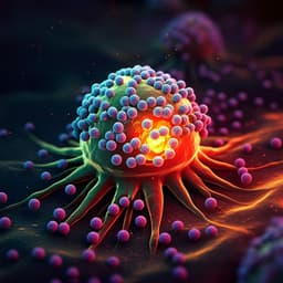
Magnetic Hybrid Nanostructures for Cancer Theranostics: A Comprehensive Review
B. Govindan, M. A. Sabri, et al.
Explore the groundbreaking research by B Govindan, M A Sabri, A Hai, F Banat, and M A A Haija, delving into magnetic hybrid nanostructures (MHNs) revolutionizing cancer theranostics. From diagnosis to therapy, discover how these innovative structures are at the forefront of magnetic resonance imaging, drug delivery, and the integration of AI to enhance treatment outcomes.
Related Publications
Explore these studies to deepen your understanding
Adjacent work that informs or extends this paper's methodology and findings.
Medicine and Health
Recent progress in the development of nanomaterials targeting multiple cancer metabolic pathways: a review of mechanistic approaches for cancer treatment
L. Zhang, B. Zhai, et al.
Medicine and Health
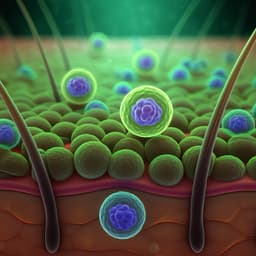
Advances in Photodynamic Therapy for the Treatment of Actinic Keratosis and Nonmelanoma Skin Cancer: A Narrative Review
A. S. Farberg, W. Justin, et al.

Medicine and Health
Plant-Based Nutrition: Exploring Health Benefits for Atherosclerosis, Chronic Diseases, and Metabolic Syndrome—A Comprehensive Review
H. Peña-jorquera, V. Cid-jofré, et al.

Medicine and Health
Nutritional Interventions during Chemotherapy for Pancreatic Cancer: A Systematic Review of Prospective Studies
M. Cintoni, F. Grassi, et al.


