PhysicsLight: Science & Applications
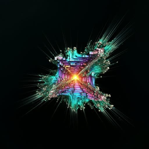
Low-dose real-time X-ray imaging with nontoxic double perovskite scintillators
W. Zhu, W. Ma, et al.
Explore the exciting advancements in X-ray imaging with innovative nontoxic double-perovskite scintillators, demonstrated by researchers Wenjuan Zhu, Wenbo Ma, Yirong Su, and others. These materials optimize absorption and emission efficiency, achieving impressive results such as a light yield surpassing CsPbBr₃, all while minimizing self-absorption. Uncover the potential for stable and high-performance imaging at low doses, even after exposure to thermal and X-ray irradiation.
Related Publications
Explore these studies to deepen your understanding
Adjacent work that informs or extends this paper's methodology and findings.
Engineering and Technology
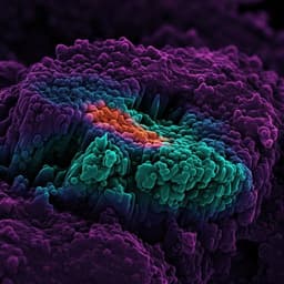
Bottom-up construction of low-dimensional perovskite thick films for high-performance X-ray detection and imaging
S. Dong, Z. Fan, et al.
Physics
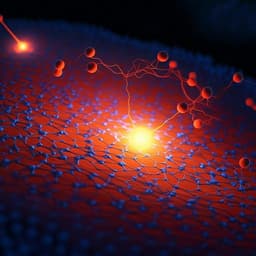
Mixed-state electron ptychography enables sub-angstrom resolution imaging with picometer precision at low dose
Z. Chen, M. Odstrcil, et al.

Engineering and Technology
Enhanced detection of threat materials by dark-field x-ray imaging combined with deep neural networks
T. Partridge, A. Astolfo, et al.
Biology
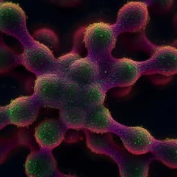
Multielement Z-tag imaging by X-ray fluorescence microscopy for next-generation multiplex imaging
M. Strotton, T. Hosogane, et al.


