
PhysicsNature Communications
In situ electron paramagnetic resonance spectroscopy using single nanodiamond sensors
Z. Qin, Z. Wang, et al.
Discover the groundbreaking research by Zhuoyang Qin and colleagues on zero-field EPR spectroscopy utilizing nanodiamonds to unravel molecular dynamics in living cells. This innovative approach presents robust spectra that promise to transform *in vivo* EPR analysis.
Related Publications
Explore these studies to deepen your understanding
Adjacent work that informs or extends this paper's methodology and findings.
Medicine and Health
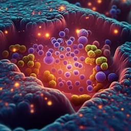
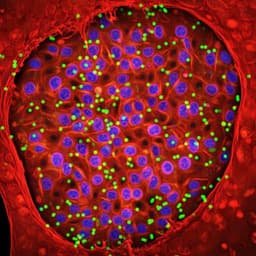
High resolution mapping of the tumor microenvironment using integrated single-cell, spatial and in situ analysis
A. Janesick, R. Shelansky, et al.

Chemistry
Mechanical cleaning of graphene using in situ electron microscopy
P. Schweizer, C. Dolle, et al.
Medicine and Health
Image-seq: spatially resolved single-cell sequencing guided by in situ and in vivo imaging
C. Haase, K. Gustafsson, et al.

Physics
High-fidelity single-shot readout of single electron spin in diamond with spin-to-charge conversion
Q. Zhang, Y. Guo, et al.


