
Medicine and Health
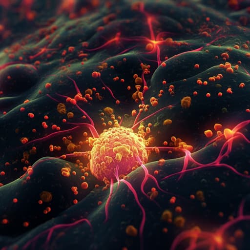
IL-15 armoring enhances the antitumor efficacy of claudin 18.2-targeting CAR-T cells in syngeneic mouse tumor models
H. Shi, A. Li, et al.
Discover how IL-15-armored claudin 18.2-targeting CAR-T cells are revolutionizing antitumor therapy in pancreatic and melanoma models. This groundbreaking study led by Hongtai Shi, Andi Li, Zhenyu Dai, and others showcases enhanced in vitro expansion and surprising tumor angiogenesis, indicating a promising direction for solid tumor treatments.
~3 min • Beginner • English
Introduction
Gastric cancer and pancreatic adenocarcinoma carry poor prognoses and limited durable responses to current therapies, underscoring the need for novel treatments. Claudin 18.2 (CLDN18.2) is highly expressed in gastric and pancreatic tumors with restricted normal tissue expression, making it an attractive target for chimeric antigen receptor T (CAR-T) therapy. While CLDN18.2-targeting CAR-T cells have shown promising response rates, durability remains limited in solid tumors. The study investigates whether armoring CLDN18.2-targeting CAR-T cells with interleukin-15 (IL-15) enhances their antitumor efficacy and persistence in immunocompetent, syngeneic mouse models, aiming to overcome key barriers in solid tumor CAR-T therapy such as limited expansion, persistence, and tumor infiltration.
Literature Review
Prior work has validated CLDN18.2 as a therapeutic target in gastrointestinal cancers, including the clinical success of the mAb zolbetuximab and early clinical responses with CLDN18.2 CAR-T cells (e.g., CT041). However, durability in solid tumors is limited. Cytokine armoring strategies (e.g., IL-7/CCL21; IL-12; IL-15) have improved CAR-T expansion, persistence, and function in several preclinical models. IL-15, a pleiotropic cytokine supporting T and NK cell survival and memory formation, has enhanced CAR-T engraftment and antitumor activity across blood and solid tumor models, though systemic IL-15 can cause toxicity. Recent mouse studies indicate IL-15 armoring improves effector function, engraftment, and TME remodeling in immunocompetent settings, providing rationale to test IL-15-armored CLDN18.2 CAR-T in syngeneic models.
Methodology
- CAR design: Second-generation murine CAR using scFv from anti-CLDN18.2 clone 69H9, murine CD8α hinge/TM, 4-1BB costimulatory domain, and CD3ζ signaling domain. Two constructs: H9 CAR (no cytokine) and H9 CAR-IL15 (C-terminal murine IL-15 linked via P2A). A non-targeting PG CAR-IL15 recognizing P329G IgG Fc served as control in some studies.
- Vector production: Retroviral vectors (MSCV) produced by 293T transfection with pCL-Eco; virus concentrated with PEG8000 and stored at −80°C.
- T-cell preparation: Primary splenic CD3+ T cells from CD45.1 C57BL/6 mice isolated (EasySep), activated with plate-bound anti-CD3 plus soluble anti-CD28 and IL-2, transduced on RetroNectin, and expanded in IL-2-containing medium. CAR expression, phenotype (CD4/CD8, CD44/CD62L, exhaustion markers), viability (AO/PI), and IL-15/IL-15Rα/pSTAT5 assessed by flow cytometry and ELISA.
- Cell lines: Mouse Panc02 (pancreatic), B16F10 (melanoma), Hepa1-6 (hepatocellular), and 293T. Panc02 and B16F10 were engineered to stably express murine CLDN18.2 or CLDN18.1 along with luciferase-GFP; luciferase used for cytotoxicity assays.
- In vitro assays: Antigen-specific cytokine release (IFN-γ, TNF-α, IL-2) via Bio-Plex after 24 h coculture at 1:1 E:T; standard 24 h luciferase-based cytotoxicity assays across E:T ratios; real-time serial dynamic killing using xCELLigence RTCA across multiple rounds without E:T readjustment.
- IL-15 quantification: ELISA for intracellular and secreted murine IL-15 from CAR-T lysates, after CD3/CD28 restimulation, after antigen stimulation, and from Hepa1-6 infected with CAR vectors.
- In vivo efficacy: Immunocompetent CD45.2 C57BL/6 mice received s.c. Panc02-CLDN18.2 (4×10^6) or B16F10-CLDN18.2 (4×10^5). Treatments: single i.v. dose of CAR-T (2.5×10^6 or 5×10^6) from CD45.1 donors with or without cyclophosphamide (CPA, 150 mg/kg i.p.) 1 day prior. Tumors measured twice weekly; tumor volume V=(L×W^2)/2; tumor growth inhibition (TGI)=1−mean(TV_treat)/mean(TV_control). Survival followed in selected experiments.
- In vivo CAR-T detection: Absolute counts in blood and spleen by flow cytometry with counting beads; composition (CD4/CD8), memory phenotypes (CD44/CD62L), and host T-cell changes.
- Tumor IHC: Formalin-fixed paraffin-embedded sections stained for CD3 (T cells), CD31 (angiogenesis), and claudin 18 (antigen). Quantified CD3 density, CLDN18 positivity, and CD31+ vessel counts.
- Statistics: Mean±SD; unpaired two-tailed t-test, one-/two-way ANOVA with multiple-comparisons; Kaplan–Meier and log-rank test for survival; significance thresholds *, P<0.05; **, P<0.01; ***, P<0.001; ****, P<0.0001.
Key Findings
- CAR-T generation and phenotype: Similar transduction efficiency for H9 CAR vs H9 CAR-IL15 (mean CAR+ 50.6% vs 46.1%, n=4). IL-15 armoring increased in vitro expansion (mean fold expansion 339.3 vs 263.9, n=3, P<0.01) and viability (88.0% vs 83.8%, n=3, P<0.001). H9 CAR-IL15 T cells showed higher central memory (TCM) fractions in CD4 and CD8 subsets (CD4: 43.4% vs 36.5%; CD8: 48.7% vs 40.0%; n=4, P<0.05) and lower expression of exhaustion markers TIM-3 and TIGIT.
- IL-15 biology: Little secreted IL-15 detected in culture supernatants; significant intracellular IL-15 detected in H9 CAR-IL15 lysates, consistent with sequestration by IL-15Rα. Increased pSTAT5 observed in CAR+ H9 CAR-IL15 cells, indicating active IL-15 signaling. IL-15 secretion was notable after CD3/CD28 stimulation but only moderate after antigen stimulation.
- In vitro function: Both CAR formats exhibited antigen-specific cytokine production (IL-2, IFN-γ, GM-CSF) and cytotoxicity against CLDN18.2+ targets (Panc02 and B16F10) with no activity against CLDN18.1+ cells. Short-term 24 h killing showed no appreciable difference between H9 CAR and H9 CAR-IL15. Serial dynamic killing revealed slightly stronger and faster killing by H9 CAR-IL15, especially in the second round, though not sustained beyond subsequent rounds.
- In vivo efficacy without lymphodepletion (Panc02-CLDN18.2): H9 CAR-IL15 significantly reduced tumor growth vs H9 CAR (Day 15 mean tumor volume 787.1 vs 1,508.0 mm^3; TGI 40.8% vs −13.5%; n=5; P<0.01), whereas H9 CAR showed no control vs UN-T.
- In vivo efficacy with CPA (Panc02-CLDN18.2): Both CARs benefited from CPA, with superior control by H9 CAR-IL15 (Day 21 mean tumor volume 30.8 vs 351.8 mm^3; TGI 91.2% vs 40.9%; n=5; P<0.0001). Two of five achieved complete regression by Day 21; one recurred by Day 27 with loss of CLDN18.2.
- In vivo efficacy with CPA (B16F10-CLDN18.2): H9 CAR-IL15 outperformed H9 CAR (Day 15 mean tumor volume 525.5 vs 1,095.3 mm^3; TGI 79.3% vs 57.0%; n=5; P<0.0001).
- Dose exploration with CPA (Panc02-CLDN18.2): At 2.5×10^6 cell dose, H9 CAR-IL15 remained superior (Day 21 mean tumor volume 51.3 vs 221.0 mm^3, P<0.0001; TGI 90.5% vs 55.8%; n=6). Survival improved with 66.7% (4/6) alive at Day 60 and significantly better overall survival vs control.
- Mechanism: In vivo, H9 CAR-IL15 promoted greater expansion of adoptively transferred CAR-T cells in blood and spleen, with higher TCM and lower TEM proportions. IHC at Day 8 showed significantly increased CD3+ T-cell infiltration and near-complete loss of CLDN18 antigen in H9 CAR-IL15 tumors, indicating rapid antigen-positive tumor cell clearance. Unexpectedly, increased CD31+ angiogenic cells were observed in H9 CAR-IL15 tumors, suggesting IL-15-related vascular changes may facilitate T-cell infiltration.
- Safety: No significant body weight loss or histopathological organ damage observed in H9 CAR-IL15–treated mice across experiments. Non-targeting PG CAR-IL15 showed no significant toxicity and only a non-significant trend to slower tumor growth, consistent with modest IL-15 single-agent effects.
Discussion
The study demonstrates that IL-15 armoring enhances CLDN18.2 CAR-T performance against solid tumors in immunocompetent mice, addressing limitations of persistence and infiltration that hinder durability. IL-15 expression improved T-cell fitness, maintaining a less differentiated, less exhausted phenotype with higher central memory content, translating to better in vitro expansion and sustained serial killing capacity. In vivo, IL-15–armored CAR-T cells expanded more robustly, infiltrated tumors more effectively, and induced rapid clearance of antigen-positive cells, yielding markedly improved tumor control in two syngeneic models, both with and without lymphodepletion. The data suggest IL-15–mediated autocrine signaling supports CAR-T survival and function, as evidenced by pSTAT5 activation and memory skewing. Additionally, the observation of increased intratumoral CD31+ vasculature raises the possibility that IL-15 may promote angiogenesis or vascular normalization that facilitates immune cell trafficking, thereby contributing to efficacy. Antigen-loss relapses indicate selective pressure by potent CAR-T activity and highlight the need to elicit broader host immunity or use multi-target strategies. Overall, IL-15 armoring appears to enhance key determinants of solid tumor CAR-T efficacy: in vivo expansion, persistence, and tumor access.
Conclusion
IL-15 armoring of CLDN18.2-targeting CAR-T cells enhances antitumor efficacy in syngeneic murine pancreatic and melanoma models by improving in vivo expansion, sustaining a memory-skewed phenotype, increasing intratumoral T-cell infiltration, and accelerating antigen-positive tumor cell clearance. These findings provide preclinical support for next-generation, IL-15–expressing CLDN18.2 CAR-T therapies and suggest potential feasibility even without lymphodepleting conditioning. Future work should elucidate the mechanistic links between IL-15 signaling and tumor vasculature, address antigen-loss escape (e.g., multi-antigen targeting or strategies to stimulate endogenous immunity), optimize vector design for controlled IL-15 expression, and incorporate safety switches for clinical translation.
Limitations
- Abstract not provided; conclusions inferred from results and discussion. Experimental limitations include: (1) Use of murine CARs and syngeneic mouse models—findings may not fully translate to human CAR-Ts or human TMEs; (2) Antigen-loss relapse observed, indicating limited induction of endogenous, antigen-independent antitumor immunity under current regimens; (3) IL-15 secretion was modest upon antigen stimulation, possibly due to construct design (C-terminal IL-15), and kinetics not fully characterized; (4) Serial in vitro killing advantage was modest and not sustained beyond early rounds, potentially due to suboptimal IL-15 accumulation; (5) Safety conclusions are limited to mouse studies—prior reports indicate potential toxicity of IL-15–expressing CAR-Ts in clinical contexts; (6) Mechanistic role of increased angiogenesis in efficacy remains correlative and requires further causal investigation.
Related Publications
Explore these studies to deepen your understanding of the subject.