Medicine and HealthNature Communications
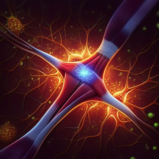
Human sensorimotor organoids derived from healthy and amyotrophic lateral sclerosis stem cells form neuromuscular junctions
J. D. Pereira, D. M. Dubreuil, et al.
This groundbreaking research showcases how human induced pluripotent stem cells (iPSC) can be utilized to model various subgroups of amyotrophic lateral sclerosis (ALS), providing insights into the nuances of disease progression through the development of functional sensorimotor organoids with neuromuscular junctions. Conducted by esteemed researchers including João D. Pereira and Brian J. Wainger, this study pushes the boundaries of precision medicine.
Related Publications
Explore these studies to deepen your understanding
Adjacent work that informs or extends this paper's methodology and findings.
Medicine and Health
Generation and characterization of cardiac valve endothelial-like cells from human pluripotent stem cells
L. Cheng, M. Xie, et al.
Medicine and Health
Injectable decellularized cartilage matrix hydrogel encapsulating urine-derived stem cells for immunomodulatory and cartilage defect regeneration
J. Zeng, L. Huang, et al.

Medicine and Health
Single-cell guided prenatal derivation of primary fetal epithelial organoids from human amniotic and tracheal fluids
M. F. M. Gerli, G. Calà, et al.

Medicine and Health
Menstrual blood-derived stromal cells modulate functional properties of mouse and human macrophages
R. Martínez-aguilar, S. Romero-pinedo, et al.


