
Medicine and Health
Human heart-forming organoids recapitulate early heart and foregut development
L. Drakhlis, S. Biswanath, et al.
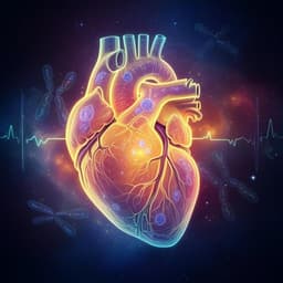
This innovative study highlights the creation of intricate, three-dimensional heart-forming organoids derived from human pluripotent stem cells, mimicking crucial heart and foregut development stages. Conducted by a team of experts, including Lika Drakhlis and Santoshi Biswanath, this research opens new pathways for understanding human heart development and related diseases.
Playback language: English
Introduction
Organoid models have revolutionized the study of various organ systems, offering unprecedented opportunities to investigate development and disease in vitro. While organoids have been successfully generated for tissues like the intestine, brain, and kidney, the cardiovascular field has lagged behind. Previous attempts to create cardiac organoids focused on engineering pre-differentiated or primary cardiac cell types to mimic adult heart tissue, neglecting the intricate processes of early heart development. This study aimed to overcome this limitation by generating HFOs that recapitulate the crucial interplay between early heart development and foregut endoderm formation. The heart is the first functional organ to form during embryogenesis, originating from the splanchnic mesoderm. This process involves the specification of precardiac mesoderm, the formation of bilateral heart-forming regions, their fusion into the heart tube, and the subsequent looping process. The interaction between the heart-forming regions and the developing foregut endoderm is essential for proper heart development. This study aimed to develop a robust method for generating human HFOs that accurately reflects these early developmental processes, creating a valuable in vitro model for studying human cardiogenesis and related diseases.
Literature Review
Existing cardiac organoid models primarily focused on adult heart tissue, using pre-differentiated cardiomyocytes or primary cells. While these models provided valuable insights, they lacked the complexity and spatiotemporal dynamics of early heart development. Studies of cardiac microtissues or organoids had improved upon previous heart muscle engineering, but these typically focused on adult-like heart tissue rather than recapitulating early development. Some studies generated cardiac microchambers using geometric confinement, or precardiac organoids from mouse PSCs, but these did not fully demonstrate the spatiotemporal patterning of early cardiogenesis and its interplay with foregut endoderm. This gap in the literature motivated the current study to develop a novel approach to recapitulate the earliest stages of human cardiogenesis in vitro.
Methodology
The researchers generated HFOs by embedding human pluripotent stem cell (hPSC) aggregates in Matrigel, followed by directed cardiac differentiation via biphasic Wnt pathway modulation using small molecules CHIR99021 (CHIR; WNT pathway activator) and IWP2 (WNT pathway inhibitor). The optimal cell number for aggregate formation was determined to be 5000 cells. The differentiation process was monitored using reporter lines expressing MIXL1-GFP (mesendoderm marker) and NKX2.5-eGFP (cardiomyogenic marker). The resulting HFOs were characterized using various techniques, including immunofluorescence (IF) staining for various markers, such as myosin heavy chain (MHC), cardiac troponin T (cTnT), Wilms' tumor 1 (WT1), vimentin, and CD31; transmission electron microscopy (TEM); scanning laser optical tomography (SLOT); multiphoton microscopy (MPM); flow cytometry; patch clamping; microarray analysis; and single-cell RNA sequencing (scRNA-seq). The researchers also generated NKX2.5-knockout (KO) HFOs to investigate the in vitro effects of NKX2.5 gene disruption. The NKX2.5-KO HFOs were compared to control HFOs using the same range of techniques. In addition, they cultured HFOs for extended periods (up to 146 days) to observe long-term morphological changes and tissue composition. They compared HFOs to conventional 2D and 3D cardiac differentiation methods using microarray analysis. For comparison with in vivo development, human embryonic hearts at 5-7 weeks of gestation (the earliest samples ethically and practically available) were analyzed alongside HFOs. The researchers used different matrices such as Matrigel, Geltrex, and collagen I to find the optimal embedding for HFOs. Statistical analysis was performed using two-tailed t-tests and one-way ANOVA.
Key Findings
The researchers successfully generated HFOs with a layered structure resembling early embryonic heart anlagen. These HFOs comprised a myocardial layer (ML) lined by endocardial-like cells, surrounded by septum transversum-like anlagen, and further contained spatially and molecularly distinct anterior versus posterior foregut endoderm tissues and a vascular network. The ML, primarily composed of second heart field (SHF)-like cells, was confirmed by immunofluorescence staining for MHC and cTnT, and electrophysiological analysis demonstrated mainly an immature ventricular-like phenotype. The outer layer contained WT1-positive cells, suggesting the presence of septum transversum-like cells. A vascular network was observed in the inner core (IC) using CD31 staining and TEM analysis, with a distinct EC layer lining the ML, possibly representing endocardium anlagen. The IC contained anterior foregut endoderm (AFE), indicated by SOX2 expression, while the outer layer expressed HNF4α, a marker for posterior foregut endoderm (PFE), specifically liver anlagen. Microarray and scRNA-seq analyses confirmed the distinct anterior-posterior endoderm patterning, identifying distinct cell populations representing cardiomyocytes, mesenchymal cells, endothelial cells, pluripotent cells, AFE, and liver anlagen/PFE. NKX2.5-KO HFOs exhibited a phenotype consistent with cardiac malformations observed in NKX2.5-deficient mice: decreased cardiomyocyte adhesion, hypertrophy, and disorganized sarcomeres. The NKX2.5-KO HFOs displayed a significantly larger total area and reduced myocardial layer compactness compared to control HFOs. However, contractility appeared to be unaffected, indicating that NKX2.5 is not essential for contractility. Microarray analysis of NKX2.5-KO HFOs revealed downregulation of NKX2.5 target genes NPPA and IRX4 and upregulation of the smooth muscle myosin gene MYH11 and NR4A3, a nuclear hormone receptor associated with cardiac hypertrophy. Long-term culture (up to 146 days) revealed that HFOs shrank, changed shape, and showed dominance of NKX2.5-positive cells over time, suggesting that the culture conditions were not optimal for long-term maintenance of all tissue components. Matrigel was found to be indispensable for the formation of layered organoids, indicating that some unidentified factors in Matrigel play a crucial role in HFO patterning.
Discussion
The HFOs generated in this study provide a powerful new in vitro model to investigate human cardiogenesis and related diseases. The recapitulation of key features of early heart and foregut development, including the intricate interplay between these two systems, makes this model unique and highly valuable. The findings demonstrate that this organoid model accurately reflects several aspects of the in vivo developmental process, such as the layered structure of the early heart anlage, the formation of endocardial-like cells, the presence of septum transversum-like cells, the distinct anterior-posterior patterning of foregut endoderm, and the development of a vascular network. The successful application of this model to study the phenotype of NKX2.5-deficient HFOs highlights the potential of this model for studying the effects of genetic mutations on early heart development. The ability to observe the long-term behavior of HFOs also offers opportunities to investigate the effects of various factors on the long-term survival and maturation of the organoids and their constituent cell types. The similarities and differences between the HFO model and in vivo development, particularly with respect to the incomplete patterning of the heart anlagen and the absence of a proper anterior-posterior axis, are noted and discussed as areas of improvement for future studies.
Conclusion
This study successfully generated a novel and robust in vitro model, the heart-forming organoid, which recapitulates key aspects of early human cardiogenesis and foregut development. These organoids exhibit striking morphological and molecular similarities to the developing heart and foregut in early human embryos, providing a valuable tool for studying developmental processes and disease modeling. Further refinements of culture conditions are suggested to promote long-term survival and maturation of all tissue compartments within the organoid, and the model has the potential to be adapted for high-throughput screening to accelerate drug discovery and investigate mechanisms of human embryogenesis. The demonstration of NKX2.5-KO phenotype recapitulation further emphasizes the potential of this model for disease modeling. Future research directions include optimizing the culture conditions to promote maturation of the HFOs and the investigation of further developmental aspects, including the formation of cardiac jelly and the role of signaling pathways. The development of a high-throughput screening platform based on HFOs would also significantly advance its applications.
Limitations
The HFOs, while closely resembling early heart and foregut anlagen, do not perfectly replicate all aspects of in vivo development. The organoids do not form a proper anterior-posterior axis, with heart anlage positioned between AFE and PFE, differing from the ventral position in embryos. Cardiac jelly is also absent from HFOs. Furthermore, the current culture conditions are not ideal for long-term maintenance of all tissue components, as evidenced by the shrinkage and shift in cell composition during prolonged culture. The limited availability of early human embryonic heart samples for comparison also poses a limitation. The success of HFO formation is dependent on the use of Matrigel, the exact components of which remain undefined, limiting the complete controllability of the experimental conditions.
Related Publications
Explore these studies to deepen your understanding of the subject.