Medicine and HealthNature Communications
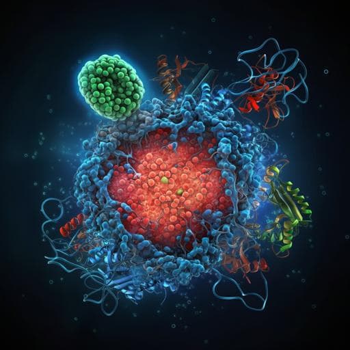
Hsc70 promotes anti-tumor immunity by targeting PD-L1 for lysosomal degradation
X. Xu, T. Xie, et al.
This groundbreaking research by Xiaoyan Xu and colleagues reveals how Hsc70 can significantly enhance the efficacy of immune checkpoint therapies by promoting the degradation of PD-L1. By inhibiting the CMTM6-PD-L1 interaction, Hsc70 plays a crucial role in reducing tumor growth and improving anti-tumor immunity, particularly when paired with the Hsp90α/β inhibitor AUY-922.
Related Publications
Explore these studies to deepen your understanding
Adjacent work that informs or extends this paper's methodology and findings.
Medicine and Health
NEK2 inhibition triggers anti-pancreatic cancer immunity by targeting PD-L1
X. Zhang, X. Huang, et al.
Medicine and Health
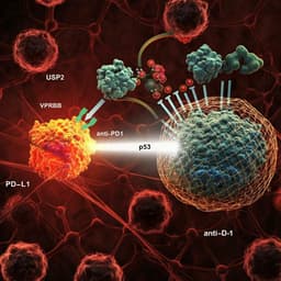
Targeting USP2 regulation of VPRBP-mediated degradation of p53 and PD-L1 for cancer therapy
J. Yi, O. Tavana, et al.
Medicine and Health
CD317 maintains proteostasis and cell survival in response to proteasome inhibitors by targeting calnexin for RACK1-mediated autophagic degradation
J. Cheng, G. Zhang, et al.
Medicine and Health
Targeting IL-21 to tumor-reactive T cells enhances memory T cell responses and anti-PD-1 antibody therapy
Y. Li, Y. Cong, et al.


