Engineering and TechnologyMicrosystems & Nanoengineering
Highly stable integration of graphene Hall sensors on a microfluidic platform for magnetic sensing in whole blood
N. Shah, V. Iyer, et al.
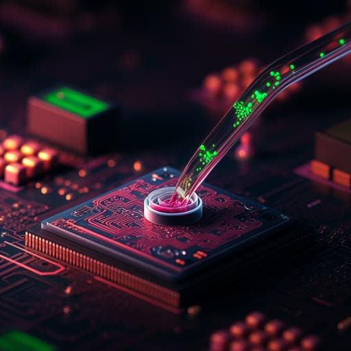
This paper presents innovative CMOS-compatible graphene Hall sensors integrated with PDMS microfluidics for magnetic sensing in blood. The developed graphene micro-Hall detectors demonstrate exceptional robustness and performance, paving the way for groundbreaking clinical applications, as validated by tests with magnetic beads by authors Nishal Shah, Vasant Iyer, Zhiping Zhang, Zhaoli Gao, Juhwan Park, Venkata Yelleswarapu, Firooz Aflatouni, A. T. Charlie Johnson, and David Issadore.
Related Publications
Explore these studies to deepen your understanding
Adjacent work that informs or extends this paper's methodology and findings.
Medicine and Health
A 3D-printed magnetic digital microfluidic diagnostic platform for rapid colorimetric sensing of carbapenemase-producing Enterobacteriaceae
P. Kanitthamniyom, P. Y. Hon, et al.
Medicine and Health
Identification of a highly stable bioactive 3-hydroxyproline-containing tripeptide in human blood after collagen hydrolysate ingestion
Y. Taga, Y. Iwasaki, et al.

Engineering and Technology
A double-layered liquid metal-based electrochemical sensing system on fabric as a wearable detector for glucose in sweat
X. Chen, H. Wan, et al.

Environmental Studies and Forestry
A new scheme for low-carbon recycling of urban and rural organic waste based on carbon footprint assessment: A case study in China
K. Zhou, Y. Li, et al.


