Medicine and Health
Function-adaptive clustered nanoparticles reverse *Streptococcus mutans* dental biofilm and maintain microbiota balance
F. Ostadhossein, P. Moitra, et al.
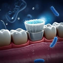
The oral cavity hosts a complex microbiota where ecological imbalance can lead to dental caries and other systemic diseases. Dental plaques are structured biofilms with an extracellular polymeric substance (EPS) that protects embedded bacteria, notably Streptococcus mutans, whose acid production (pH ~4.5) demineralizes enamel. Conventional approaches (mechanical removal, fluoride, chlorhexidine/CHX) have limitations including disputed efficacy, side effects, and risk of resistance. Nanoparticles offer multifunctionality and stimulus-responsiveness, but many fail against mature biofilms or pose biodegradability concerns. The research question is whether pH-responsive, antibiotic-free, clustered carbon dot nanoparticles (CDots) can eradicate mature S. mutans biofilms by activating within acidic biofilm microenvironments while preserving overall oral microbiota balance. The study proposes a particle-in-particle design: phosphonium-functionalized therapeutic CDots encapsulated within a pH-responsive PS-b-PDMA shell to enable contact-mediated killing in acidic biofilm niches.
Prior work identifies S. mutans as a dominant cariogenic pathogen in biofilms and highlights EPS and low pH as key virulence factors. CHX is widely used but with debated antiplaque/anticaries efficacy and notable side effects (staining, tartar, dysgeusia). Traditional antibiotics risk resistance and can disrupt beneficial commensals. Nanoparticle strategies provide controlled release, selectivity, and imaging, with activatable designs reducing off-target effects; however, concerns exist regarding metallic NP persistence and limited efficacy against mature biofilms. Phosphonium-containing antimicrobials can outperform ammonium analogs. Carbon dots offer facile synthesis, luminescence, functionalizable surfaces, and degradability, making them promising antibacterial platforms. Prior NP approaches to oral biofilms often require external stimuli (e.g., H2O2, light) or drugs; a drug-free, biofilm pH-activated system could mitigate toxicity and resistance while maintaining microbiome homeostasis.
Nanoparticle synthesis and characterization: Carbon dot nanoparticles (CDots) were synthesized hydrothermally (180 °C, ~2 MPa, 24 h) from chlorhexidine (CHX) alone (CHX NPs) or CHX admixed with 30 wt% tributylhexadecylphosphonium bromide (PR4+) (CHX PR4+ NPs) in methanol. Post-synthesis processing included sonication, filtration (0.45/0.22 µm), solvent removal, and drying. For pH-responsive encapsulation, PS-b-PDMA was co-dissolved with CDots in CHCl3, thin-film formed by N2 evaporation, residual solvent removed (vacuum), then hydrated in water to yield polymer-wrapped nanoparticles (CHX polymer NPs, CHX PR4+ polymer NPs). Empty micelles were similarly prepared. Physicochemical analyses: TEM measured anhydrous diameters (CHX PR4+ NPs ~2.6±0.6 nm; CHX PR4+ polymer NPs ~26±4 nm). Hydrodynamic sizes and stability were assessed by nanoparticle tracking analysis (ZetaView). AFM confirmed increased height after wrapping. Fluorescence spectroscopy verified maintained/enhanced CDot emission upon encapsulation. Zeta potential: CHX polymer NPs −13±3 mV; CHX PR4+ polymer NPs +35±4 mV. pH-responsive release studied via dialysis in PBS (pH 7.3) vs acetate buffer (pH 4.5) at 37 °C with UV–vis quantification showed faster release under acidic conditions. Chemical structure/provenance assessed by MS (loss of CHX parent peak m/z 505.3 in CDots), 1H/31P NMR (phosphorus retained), and XPS (P 2p doublets at 132.3/133.1 eV; Cl binding energy shifts), indicating carbonization altered CHX while preserving phosphonium features. In vitro antibacterial assays (planktonic S. mutans): Turbidity time-kill (OD600) recorded every 30 min; minimum inhibitory concentration (MIC) determined using Lambert and Pearson’s model. Live/dead staining with SYTO9/PI quantified viability at 0.7–5.7 mM. Intracellular ROS measured with DCFH-DA. Membrane potential assessed by DiOC2(3) ratiometric red:green flow cytometry. SEM/TEM visualized membrane integrity and NP attachment. DNA damage evaluated by agarose gel electrophoresis of plasmid pBR322 and extracted genomic DNA, and by TUNEL (Tunnelyte Red) confocal imaging with Hoechst nuclear counterstain. In vitro biofilm assays: 48-h mature S. mutans biofilms formed in 96-well plates. Dispersion assessed by 0.5% crystal violet and resazurin assays after 4 h treatment across concentrations. Inhibition assessed by preincubating bacteria with NPs during biofilm formation (≈52 h) followed by resazurin readout. CHX comparisons in some assays were limited by CHX aqueous solubility. Ex vivo human tooth model: Sterilized extracted molars were embedded on agarose supports, inoculated with S. mutans (OD600=0.6), incubated 48 h to form biofilm, treated with NPs (5.7 mM, 4 h), then gently sonicated to recover cultivable bacteria for colony counting on BHI agar. Degradation in artificial saliva: CHX PR4+ NPs (5.7 mM) incubated in artificial saliva (pH 6.8, 37 °C). UV–vis absorbance at 253 nm tracked over 10 days; ESI–MS profiled degradation intermediates and proposed pathways from carbonized CHX to smaller metabolites. In vivo rat model: Female Sprague Dawley rats (n=3/group) prescreened S. mutans-free, divided into water, CHX, and CHX PR4+ polymer NP groups. S. mutans inoculation on incisors for 6 days, 7-day establishment, then topical treatments daily (1.4 mM, 100 µl, 1 min) for 11 days, followed by 2 days at 2.3 mM. S. mutans presence assessed by lateral flow kit pre-sacrifice. Teeth harvested; bacteria dislodged and plated on mitis salivarius-bacitracin agar selective for S. mutans to enumerate CFU. Safety evaluated by histopathology (H&E) of major organs and gingiva. Microbiota analysis: Post-treatment dental swabs underwent DNA extraction and 16S rRNA gene amplicon sequencing (V3–V4) on Illumina MiSeq. Data processed via dada2/DECIPHER; alpha diversity (Shannon, Chao1), beta diversity (Bray-Curtis, PCoA, PERMANOVA, ANOSIM), and taxonomic composition compared across groups. Statistics performed in GraphPad Prism; results presented as mean±SD with ANOVA where applicable.
- Nanoparticle platform: Phosphonium-functionalized CDots encapsulated in PS-b-PDMA formed clustered "particle-in-particle" NPs (~26±4 nm by TEM; CHX PR4+ cores ~2.6±0.6 nm). Zeta potentials indicated successful phosphonium incorporation (+35±4 mV for CHX PR4+ polymer NPs). Encapsulation preserved/enhanced fluorescence.
- pH-responsive release: Faster cargo release at acidic pH (acetate buffer, ~4.5) than neutral PBS, aligning with cariogenic biofilm microenvironments.
- Planktonic antibacterial efficacy: Time-kill turbidity assays showed continuous viability decrease with CHX PR4+ NPs and CHX PR4+ polymer NPs; CHX NPs without phosphonium were ineffective. MIC for CHX PR4+ systems was 5.7 mM (calculated using NP active component molar mass of 355 g/mol). Live/dead assays showed markedly higher dead-cell percentages for CHX PR4+ polymer NPs versus controls and CHX NPs. ROS levels were significantly elevated with CHX PR4+ polymer NPs, exceeding CHX positive control. Membrane depolarization occurred: DiOC2(3) red:green ratio dropped from ~467 (control) to ~204 (NP-treated), indicating loss of membrane potential.
- Mechanism: SEM/TEM revealed extensive membrane damage and numerous NPs attached to S. mutans surfaces, with severe cellular deformation/collapse. DNA damage evidenced by complete plasmid degradation at 5.7 mM, genomic DNA fragmentation (bands migrating beyond ladder), and increased TUNEL positivity (p=0.001 vs water), indicating DNA nicking/fragmentation.
- Biofilm effects in vitro: CHX PR4+ polymer NPs dispersed 48-h mature biofilms in a dose-dependent manner (crystal violet, resazurin). Biofilm formation was inhibited by >90% at 0.7–5.7 mM during preincubation (resazurin).
- Ex vivo human tooth model: Treatment (5.7 mM, 4 h) significantly reduced cultivable bacteria recovered from tooth surfaces compared with water control, corroborated by SEM showing depleted biofilm and NP-bacteria interactions.
- Degradability: In artificial saliva, NP absorbance decreased over 10 days; ESI–MS detected progressive formation of smaller intermediates (e.g., m/z 427, 637, 704, 705, 722, 740, 741, 755; later 680, 781; further 686, 701, 723, 743; eventually 773, 828), supporting biodegradation to smaller metabolites.
- In vivo efficacy: In a rat model, CHX PR4+ polymer NP treatment yielded negative S. mutans lateral flow tests akin to CHX, while water controls were positive. Plate counts of cultivable S. mutans from teeth were significantly lower with CHX PR4+ polymer NPs versus water (p=0.0008) and superior to CHX (p=0.0308 w.r.t. water). No animal loss and similar weight gain across groups; gingival histology indicated minimal-to-no lesions in NP group compared with mild changes in controls; no significant organ histopathology differences.
- Microbiota preservation: 16S rRNA sequencing showed no significant differences across groups in alpha diversity (Shannon p=0.204; Chao1 p=0.341) or beta diversity (Bray-Curtis PERMANOVA p=0.16; ANOSIM p=0.294). Taxonomic composition proportions were similar among groups, indicating maintained microbial richness and diversity after NP treatment.
- Overall: The pH-activated, antibiotic-free, phosphonium-CDot clustered NPs eradicated S. mutans and disrupted biofilms (>90% inhibition in vitro), suppressed biofilm in vivo, showed biodegradability in saliva simulant, and preserved oral microbiota diversity.
Targeting the acidic microenvironments within dental biofilms enabled on-site activation and release of clustered therapeutic CDots, producing strong contact-mediated interactions with S. mutans membranes, elevated ROS, membrane depolarization, and DNA fragmentation. This multi-pronged mechanism translated into robust inhibition and dispersion of mature biofilms in vitro and on human tooth surfaces ex vivo, and into significant suppression of S. mutans biofilm burden in vivo. Crucially, the strategy spared the broader oral microbiota, avoiding reductions in richness/diversity and large compositional shifts, addressing a common drawback of broad-spectrum antiseptics like CHX. The absence of an external stimulus requirement (e.g., H2O2 or light) reduces potential off-target toxicity, and degradability in artificial saliva suggests favorable clearance/metabolism profiles relative to persistent metallic NPs. Collectively, findings demonstrate that a pH-responsive particle-in-particle platform can selectively target cariogenic conditions, mitigate dental biofilms, and maintain ecological balance, aligning with precision antibiofilm goals and potentially lowering resistance risks by using an antibiotic-free, physically/chemically driven mechanism.
This work introduces a pH-responsive, clustered carbon dot nanoparticle system that effectively disrupts and inhibits S. mutans biofilms while preserving oral microbiota diversity and richness. The platform leverages acidic biofilm niches for on-site activation, achieving >90% biofilm inhibition in vitro, significant suppression in vivo, and evidence of biodegradation in saliva simulant. The antibiotic-free, stimuli-free approach offers a promising alternative to conventional antiseptics, with potential for reduced side effects and resistance. Future directions include: optimizing dosing/formulations (e.g., oral pastes/rinses), expanding to multispecies biofilm models and clinical isolates, integrating active targeting ligands for specific pathogens, comprehensive long-term safety and pharmacokinetic studies, and translation to other biofilm-driven infections (e.g., periodontitis, implant-associated biofilms, endocarditis).
- Some comparative experiments with CHX were limited by its poor aqueous solubility, restricting direct head-to-head assessments in certain biofilm assays.
- In vivo studies used small group sizes (n=3 per group), warranting larger cohorts for robust statistical power and evaluation of variability.
- Cytotoxicity assessments in NIH 3T3 cells indicated dose-dependent ROS and reduced viability at higher NP concentrations, necessitating careful dose optimization for clinical translation.
- The primary in vitro pathogen was S. mutans; while relevant, broader testing against polymicrobial cariogenic communities and mixed-species biofilms would strengthen generalizability.
- Degradation studies were performed in artificial saliva; in vivo degradation, clearance, and metabolite safety profiles require further validation.
Related Publications
Explore these studies to deepen your understanding of the subject.