
Medicine and HealthNature Communications
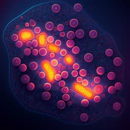
Full spectrum fluorescence lifetime imaging with 0.5 nm spectral and 50 ps temporal resolution
G. O. S. Williams, E. Williams, et al.
This paper introduces a groundbreaking high-speed optical scanning microscope that captures time-resolved images across 512 spectral and 32 time channels within a single acquisition, providing insights into complex organic structures. The research by Gareth O. S. Williams and colleagues demonstrates its potential through imaging various samples, including human lung tissue, revealing intricate spectral changes.
Related Publications
Explore these studies to deepen your understanding
Adjacent work that informs or extends this paper's methodology and findings.

Biology
Fluorescence lifetime imaging with a megapixel SPAD camera and neural network lifetime estimation
V. Zickus, M. Wu, et al.
Biology
Ultra-high spatio-temporal resolution imaging with parallel acquisition-readout structured illumination microscopy (PAR-SIM)
X. Xu, W. Wang, et al.

Engineering and Technology
Multiplexed fluorescence and scatter detection with single cell resolution using on-chip fiber optics for droplet microfluidic applications
P. Gupta, A. Mohan, et al.

Engineering and Technology
Fast non-line-of-sight imaging with high-resolution and wide field of view using synthetic wavelength holography
F. Willomitzer, P. V. Rangarajan, et al.


