
Food Science and TechnologyNature Communications
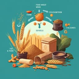
Food amyloid fibrils are safe nutrition ingredients based on in-vitro and in-vivo assessment
D. Xu, J. Zhou, et al.
Discover the fascinating world of food protein amyloid fibrils, which exhibit remarkable properties compared to their native counterparts. This study by a team of experts investigates the safety of these fibrils, revealing that they are not only safe to consume but could also offer significant nutritional benefits.
Related Publications
Explore these studies to deepen your understanding
Adjacent work that informs or extends this paper's methodology and findings.
Food Science and Technology
Assessment and scenario hypothesis of food waste in China based on material flow analysis
S. Jiang, H. Chen, et al.

Environmental Studies and Forestry
A new scheme for low-carbon recycling of urban and rural organic waste based on carbon footprint assessment: A case study in China
K. Zhou, Y. Li, et al.
Medicine and Health
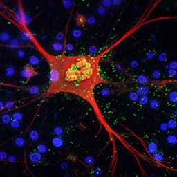
Turn-on chemiluminescence probes and dual-amplification of signal for detection of amyloid beta species in vivo
J. Yang, W. Yin, et al.
Food Science and Technology
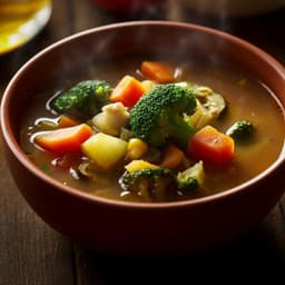
Validation of preferred salt concentration in soup based on a randomized blinded experiment in multiple regions in Japan—influence of umami (L-glutamate) on saltiness and palatability of low-salt solutions
H. Hayabuchi, R. Morita, et al.


