Fluorescence lifetime imaging with a megapixel SPAD camera and neural network lifetime estimation
V. Zickus, M. Wu, et al.
Explore these studies to deepen your understanding
Adjacent work that informs or extends this paper's methodology and findings.
Full spectrum fluorescence lifetime imaging with 0.5 nm spectral and 50 ps temporal resolution
G. O. S. Williams, E. Williams, et al.

Powering AI at the edge: A robust, memristor-based binarized neural network with near-memory computing and miniaturized solar cell
F. Jebali, A. Majumdar, et al.
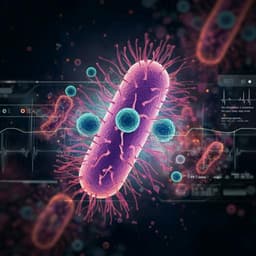
Rapid species identification of pathogenic bacteria from a minute quantity exploiting three-dimensional quantitative phase imaging and artificial neural network
G. Kim, D. Ahn, et al.

Crisis of objectivity: using a personalized network model to understand maladaptive sensemaking in a patient with psychotic, affective, and obsessive-compulsive symptoms
A. Oblak, M. Kuclar, et al.


