Medicine and Health
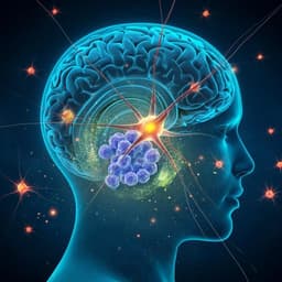
Exercise reverses learning deficits induced by hippocampal injury by promoting neurogenesis
L. N. Codd, D. G. Blackmore, et al.
Cognitive deficits commonly follow neurodegenerative diseases and brain injuries, including stroke, with stroke survivors showing acute impairments and accelerated long-term cognitive decline. Hippocampal atrophy, frequently observed post-stroke, correlates with impaired cognition. The adult hippocampus retains neurogenic capacity that contributes to contextual spatial learning and memory in mice, and stimulation of adult neurogenesis (e.g., via exercise) has been associated with improved spatial learning. However, standard rodent stroke models (e.g., MCAO) often entail short survival and motor deficits that confound long-term assessment of hippocampal-dependent learning. The authors developed a unilateral hippocampal injury model using ET-1 to produce a measurable learning deficit without motor impairment, enabling evaluation of whether voluntary exercise-induced neurogenesis can rescue hippocampal-mediated learning after injury and whether neurogenesis in one or both hippocampi is necessary/sufficient for recovery.
Prior work demonstrates that adult hippocampal neurogenesis is essential for spatial/contextual learning and memory in rodents, and that exercise enhances neurogenesis and spatial learning. Clinical and imaging studies link hippocampal atrophy after cortical/subcortical strokes with cognitive impairment. Exercise interventions in humans post-stroke have shown improved cognitive performance. Nevertheless, effects of exercise after experimental stroke on hippocampal neurogenesis have been mixed, with reports of both decreased and increased newborn neuron numbers. The commonly used MCAO model complicates cognitive assessments due to motor deficits, motivating alternative models to isolate hippocampal-dependent functions. Additional literature implicates neurotrophic and molecular mediators (e.g., BDNF, serotonin signaling, growth hormone, microRNA-124, TrkB, REST) in exercise-induced neurogenesis and cognitive recovery.
Design: Female C57BL/6 and DCXDTR mice (10–13 weeks) underwent unilateral intrahippocampal ET-1 injection (right hemisphere) to induce focal hippocampal injury; contralateral PBS served as control. Cognitive testing used the active place-avoidance (APA) task in three blocks: pre-injury (APA1), post-injury (APA2), and post-run (APA3), with visual cues changed between blocks. Some experiments extended to APA4–APA5.
ET-1 lesion: Under ketamine/xylazine anesthesia and stereotaxic guidance, 1 μL ET-1 (333 pmol; with Fluororuby) was injected into the right hippocampal fissure (AP −1.8 mm, ML ±1.0 mm, DV −1.7 mm relative to Bregma) over 5 min; contralateral PBS (1 μL) was injected similarly. Pipette was left in place to minimize backflow. Sham animals received bilateral PBS. Animals recovered with analgesia (butorphanol) and antibiotic (enrofloxacin).
Voluntary running: Mice were group-housed (3–4/cage) with angled-disc wireless running wheels for 21 days; collective cage running distance was recorded. BrdU (100 mg/kg i.p.) was administered daily during the last 4 days of running to label proliferating cells.
Neurogenesis manipulation: DCXDTR transgenic mice expressed human diphtheria toxin receptor under the DCX promoter. Systemic ablation: Intraperitoneal diphtheria toxin (DT) 10 ng/g every second day for 14 days (7 injections) starting day 31 post-ET-1 to ablate DCX+ immature neurons generated during running, followed by APA testing and tissue analysis. Local ablation: Unilateral or bilateral intrahippocampal DT injections (10 pg/0.5 μL PBS) into the hilus (AP −2.0 mm, ML ±1.5 mm, DV −2.0 mm) on the final day of running to ablate DCX+ neurons selectively in targeted hippocampi, followed by APA testing at 2 and 3+ weeks post-injury (and up to 22 weeks for bilateral injections).
Behavioral testing (APA): Mice learned to avoid a stationary shock-zone defined relative to distal visual cues on a rotating platform (1 rpm). Sessions: 1 day habituation (5 min, no shock) then 5 days of testing per block (10 min/day). Shock: 0.5 mA, 500 ms, 1.5 s intershock interval. Outcome measures: number of shocks, maximum time avoiding the shock-zone, distance traveled. Visual cues differed between APA blocks. Additional motor assessments included accelerating Rota-Rod, open-field activity monitoring (distance, velocity), and fore/hindlimb grip strength pre- and one week post-surgery.
Histology and quantification: After perfusion and fixation, coronal brain sections (40 μm) were processed for immunohistochemistry: BrdU, DCX (immature neurons), and NeuN (mature neurons), with DAPI counterstain. Seven sections per animal (spanning hippocampus) were analyzed by blinded investigators. Measures included hippocampal area, spared dentate gyrus granule cell layer (GCL) area, inner GCL length, and densities per DG mm length of DCX+, BrdU+, BrdU/DCX+, and BrdU/NeuN+ cells.
Statistics: Data are mean ± SEM. APA data analyzed via repeated-measures two-way ANOVA with Bonferroni post hoc comparisons across days and between groups (predefined comparisons). Cell counts: paired t-tests (ET-1 vs vehicle hippocampus within subject) and unpaired t-tests (between treatments/genotypes). Motor tests: paired (pre vs post) and unpaired (PBS vs ET-1) t-tests. Power calculations (α=0.05, power=0.80) informed sample sizes. Animals were grouped by similar APA performance before random assignment to conditions. APA data collection was automated; cell counts were blinded.
- ET-1 produced a unilateral hippocampal lesion predominantly affecting the dentate gyrus, with significant atrophy: total hippocampal area reduced by 18.5 ± 5.0% (paired t-test t=3.157, df=7, p=0.0160) and spared dentate GCL area reduced by 14.3 ± 3.8% (t=3.557, df=7, p=0.0093) compared with contralateral vehicle-injected hippocampus.
- Pre-injury (APA1): All groups improved over 5 days (shocks reduced; F=14.6, p<0.0001) with no group differences (F=0.1668, p=0.8470). Max time avoided increased across days (F=8.342, p<0.0001) but day 1 vs 5 post hoc not significant.
- Post-injury (APA2): ET-1–injured mice failed to learn; no within-block improvement (F=0.4157, p=0.7971). Between-group differences were significant (F=5.058, p=0.0114); ET-1 mice received more shocks than vehicle (p=0.0493) and naïve (p=0.0084) and worse than their pre-injury performance (paired t p=0.0234). Max time avoided did not differ between groups (F=1.734, p=0.1906). ET-1 did not affect distance traveled (F=2.639, p=0.0849). Motor tests showed no deficits vs PBS except higher average forelimb grip strength in ET-1 mice.
- Running behavior: ET-1–injured and vehicle mice ran similar daily distances (0.79±0.08 km vs 0.97±0.11 km; p=0.2).
- Post-run (APA3): ET-1 Injury + Run animals showed recovery, performing comparably to non-injury runners (overall F=4.112, p=0.0078). ET-1 Injury + No Run worsened (day 1 vs 5 p=0.0002) and received more shocks than all other groups on the final day (all p<0.0001). Max time avoided increased over time for all except ET-1 No Run (F=6.802, p<0.0001); ET-1 No Run was lower than others on final day (ET-1 Run p=0.0226; Vehicle No Run p=0.0002; Vehicle Run p=0.0253; Naïve p=0.0121).
- Neurogenesis with running: Runners had significantly more BrdU+ cells, BrdU/NeuN+ cells, and DCX+ cells in both hippocampi: vehicle hemisphere p=0.0015 (BrdU), 0.0181 (BrdU/NeuN), 0.0004 (DCX); ET-1 hemisphere p=0.0006 (BrdU), 0.0156 (BrdU/NeuN), 0.0485 (DCX). DCX+ neurons were reduced in ET-1 vs vehicle hippocampus in both runners (p=0.0348) and non-runners (p=0.0388). BrdU/DCX+ cells increased in vehicle hippocampus of runners vs non-runners (p=0.0292).
- Structural change: ET-1-injured hippocampal area was 19% larger in runners than non-runners (unpaired t t=2.424, df=14, p=0.0295).
- Correlations: In runners, higher BrdU+ or BrdU/NeuN+ cell densities correlated with better APA3 performance (fewer shocks) in both hemispheres (R^2≈0.58–0.67; p≈0.012–0.0286).
- Necessity of neurogenesis: Systemic DT in DCXDTR mice during the immediate post-running period ablated DCX+ cells and BrdU/DCX+ cells and abolished behavioral recovery (APA3 repeated-measures two-way ANOVA F=9.346, p=0.0085); WT controls recovered.
- Sufficiency of unilateral neurogenesis: Unilateral intrahippocampal DT injection into either the ET-1–injured or contralateral hippocampus on the final day of running prevented learning at APA3 (performance similar to WT ET-1 Injury + No Run) but animals recovered by APA4. In contrast, bilateral intrahippocampal DT injections prevented recovery at APA3 and animals failed to learn even up to 22 weeks post-injury (APA4–APA5).
The study demonstrates that voluntary exercise reverses hippocampal injury-induced spatial learning deficits through increased adult neurogenesis in the dentate gyrus. Behavioral recovery after ET-1–induced unilateral hippocampal damage coincided with elevated proliferation and maturation of newborn neurons in both hippocampi. Causality was established by ablating DCX+ immature neurons: systemic ablation after running eliminated recovery, while bilateral intrahippocampal ablation blocked recovery long-term. Critically, unilateral ablation in either hemisphere only delayed recovery, indicating that enhanced neurogenesis in a single hippocampus—whether injured or contralateral—is sufficient for functional compensation. These results suggest that recovery likely reflects compensatory integration of newborn neurons into spared dentate circuits within the lesioned hemisphere or augmented function of the contralateral hippocampus, rather than obligatory structural repair of the entire damaged DG. The observed increase in hippocampal area with running supports concomitant structural plasticity. This ET-1 model, which avoids motor confounds, permits precise assessment of hippocampal-dependent cognition and highlights activatable precursor cells as therapeutic targets. Findings align with clinical observations that exercise improves hippocampal volume and cognition in older adults and suggest that targeting molecular mediators of exercise-induced neurogenesis (e.g., BDNF, serotonin pathways) could benefit patients unable to exercise.
Voluntary running restores hippocampal-dependent spatial learning after unilateral ET-1–induced hippocampal injury by stimulating adult neurogenesis. Recovery requires newborn DCX+ neurons; bilateral ablation prevents recovery, whereas neurogenesis in either the injured or uninjured hippocampus alone is sufficient for functional rescue. The study introduces a unilateral hippocampal injury model enabling long-term cognitive assessments without motor confounds and provides causal evidence that enhancing hippocampal neurogenesis can reverse injury-induced learning deficits. Future work should assess generalizability across ages, sexes, comorbidities, and species; delineate the relative contributions of repair versus compensation; characterize circuit-level and structural changes (e.g., dendritic remodeling); and explore pharmacological or neuromodulatory strategies (e.g., BDNF/serotonin pathways) to stimulate neurogenesis in patients unable to exercise.
- Model and population: Young female mice only; results may not generalize to males, aged animals, or those with comorbidities. The ET-1 unilateral hippocampal lesion does not follow STAIR recommendations favoring MCAO models, potentially limiting translational relevance to typical stroke pathology.
- Mechanistic ambiguity: Unilateral sufficiency of neurogenesis indicates compensatory mechanisms; the study cannot confirm repair within the injured hippocampus versus compensation by the contralateral side.
- Structural analyses: While hippocampal area increased with running, detailed assessments of dendritic spine density, arborization, or white matter changes were not performed.
- Behavioral blinding: Researchers were not blinded during APA testing (though data collection was automated), which could introduce bias.
- Running quantification: Voluntary running distances were recorded per cage (group-housed), not per individual, limiting precise attribution of dose-response relationships.
- Scope of motor assessment: Motor tests were performed at one week post-surgery; subtle or long-term motor changes cannot be fully excluded (though APA distance traveled was largely unaffected).
Related Publications
Explore these studies to deepen your understanding of the subject.