
Medicine and Health
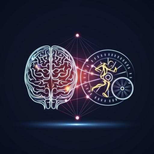
Exercise mitigates a gut microbiota-mediated reduction in adult hippocampal neurogenesis and associated behaviours in rats
S. Nicolas, S. Dohm-hansen, et al.
Discover how exercise can counteract the adverse effects of gut microbiota disruption on adult hippocampal neurogenesis and related behaviors in rats. This intriguing study by Sarah Nicolas and colleagues provides valuable insights into the connection between gut health and brain function.
~3 min • Beginner • English
Introduction
The gut microbiota, comprising trillions of microorganisms in the gastrointestinal tract, is a critical component of bidirectional gut–brain communication and has been implicated in neurological and neuropsychiatric diseases including Alzheimer's disease, Parkinson's disease, and depression. Microbiota composition varies across the lifespan and is influenced by lifestyle factors such as diet and exercise. Exercise modifies gut microbial composition and metabolite profiles, increasing diversity and taxa that produce health-promoting metabolites like short-chain fatty acids, and is associated with benefits for brain health and cognition. The hippocampus, which supports learning, memory, and mood-related behaviors, exhibits adult hippocampal neurogenesis (AHN), a plasticity process implicated in spatial/contextual memory, anxiety-like behaviors, and particularly pattern separation. Exercise robustly enhances AHN and certain cognitive functions in rodents, while the microbiome regulates cognitive behaviors, neuroplasticity, and AHN. However, how exercise interacts with microbiota disruption to influence AHN and associated behaviors remains unclear. The study investigates whether voluntary exercise can ameliorate the negative impacts of antibiotic-induced gut microbiota disruption on AHN, cognitive function, and mood-related behaviors in adult male rats.
Literature Review
Methodology
Study design: Adult (9–10 week old) male Sprague-Dawley rats (Envigo, UK) were maintained on a 12 h light/dark cycle at 22±1 °C with ad libitum chow and water. Rats were pair-housed to avoid social isolation effects on neurogenesis and randomly assigned (n=10/group) to: sedentary control (Sed), sedentary with antibiotics (Sed+ABX), voluntary running exercise (Ex), or exercise with antibiotics (Ex+ABX). An antibiotic cocktail was administered in drinking water throughout the study (bottles changed every 2 days). Two weeks after starting antibiotics, rats in Ex and Ex+ABX groups received continuous access to a running wheel (33 cm diameter; Tecniplast, UK) in dedicated cages; revolutions were continuously recorded.
Neurogenesis labeling: All rats received intraperitoneal 5-bromo-2'-deoxyuridine (BrdU; 150 mg/kg/day) for 5 consecutive days beginning two weeks after antibiotic initiation.
Behavioral testing: Commenced three weeks after exercise/BrdU initiation and continued while exercise access remained, for a total of 8 weeks of exercise. Tests (conducted during light phase with >45 min acclimation and apparatus cleaning between animals) included: modified spontaneous location recognition task (MSLRT; large and small separation conditions assessing spatial discrimination/pattern separation), two-trial Y-maze (spatial reference memory), novelty suppressed feeding test (NSFT; latency to eat and latency to approach center; post-test 30 min food intake), elevated plus maze (EPM; anxiety-like behavior), and forced swim test (FST; immobility, swimming, climbing). Scoring was blinded where possible or via automated tracking (Ethovision XT 7.0; AnyMaze).
Physiology and molecular measures: Running distance, body weight, and plasma corticosterone were measured. Skeletal muscle PGC-1α mRNA was quantified. Colon inflammatory gene expression (TNFα, IL-6, IL-10, CCL2) was assessed by qPCR. Plasma cytokines (TNFα, IL-6) were measured.
Tissue collection: At week 10, five rats/group were perfused for immunohistochemistry; brains were post-fixed, cryoprotected, sectioned coronally (40 μm; 1:12 series through hippocampus), and stored for processing. Remaining rats were decapitated; hippocampus, colon, cecal contents, and muscle were snap-frozen. Blood (EDTA) was centrifuged and plasma stored at −80 °C.
Immunohistochemistry: Adult hippocampal neurogenesis was quantified via BrdU/NeuN double-labeling in the dentate gyrus (whole, dorsal, ventral), and doublecortin (DCX) immunoreactivity with morphological sub-classification. Representative imaging at 20–40x magnification.
Metabolomics: Untargeted mass spectrometry-based metabolomics (MS-OMICS, Denmark) on cecal contents and hippocampus; 610 cecal and 210 hippocampal annotated metabolites quantified. Differential abundance analyzed with FDR correction; pathway enrichment performed. Targeted examination of TMAO, ergothioneine, carnitine/choline metabolites; assessment of antibiotic penetration into hippocampus (none detected at 10 weeks).
Statistics: Normality and variance tested (Shapiro-Wilk). Parametric data: two-way ANOVA (factors: exercise, antibiotics) with Tukey post hoc. Non-parametric: Kruskal–Wallis with Dunn’s post hoc. Significance p<0.05; metabolomics features significant at FDR-adjusted p<0.05. Spearman correlations between metabolites and biological/behavioral measures were assessed with FDR correction (5% threshold). Sample sizes noted per assay (typically n=8–10 for behavior; n=5 for IHC; see Supplementary Tables). Ethical approval: HPRA (Ireland) and University College Cork Animal Ethics; compliance with EU Directive 2010/63/EU.
Key Findings
- Body weight and running activity: Neither antibiotics nor exercise altered body weight. Exercise groups ran comparable distances across Ex and Ex+ABX. Exercise increased skeletal muscle PGC-1α expression and reduced plasma corticosterone independent of antibiotics (Sed vs Ex p=0.0327; Sed+ABX vs Ex+ABX p=0.0084).
- Peripheral and colonic inflammation/physiology: Antibiotics increased colonic TNFα mRNA and CCL2; IL-10 mRNA was increased by exercise and antibiotics individually but reduced when combined. Plasma TNFα increased with antibiotics in sedentary rats (Sed vs Sed+ABX p=0.0475). Plasma IL-6 increased only when antibiotics were combined with exercise (Ex vs Ex+ABX p=0.0063). Antibiotics tended to increase fecal output (p=0.0609) and significantly affected colon length and cecum weight (main effects of antibiotics).
- Metabolomics: Antibiotics caused a large shift in the cecal metabolome; 527 of 610 metabolites were differentially regulated in Sed+ABX vs Sed (FDR-adjusted p<0.05), with pathway enrichment in aminoacyl-tRNA biosynthesis, TCA cycle, and amino acid pathways. Exercise had minimal effects on cecal metabolites (6 in Ex vs Sed; 1 in Ex+ABX vs Sed+ABX). Ethyl 2-(4-oxo-4,5-dihydro-1,3-thiazol-2-yl)acetate was consistently decreased by exercise and increased by antibiotics. In the hippocampus, only two metabolites differed with antibiotics: TMAO decreased (PFDR=1.5E-5) and ergothioneine increased (PFDR=0.045); no metabolites were significantly altered by exercise.
- Cognitive performance: In MSLRT large separation, antibiotics impaired performance (main effect). In small separation (pattern separation), antibiotics impaired performance (Sed vs Sed+ABX p=0.0003) and exercise attenuated this deficit (Sed+ABX vs Ex+ABX p=0.045; significant Abx x Ex interaction). In the Y-maze, antibiotics reduced the discrimination ratio (main effect) without affecting locomotion.
- Anxiety- and depression-like behaviors: In NSFT, exercise reduced latency to eat (main effect), though weight loss in exercise groups may confound hunger; antibiotics increased latency to approach center (Sed vs Sed+ABX p=0.0192), which was reversed by exercise (Sed+ABX vs Ex+ABX p=0.0051). In EPM, antibiotics reduced time in open arms (Sed vs Sed+ABX p=0.0096), reversed by exercise (Sed+ABX vs Ex+ABX p=0.028). In FST, exercise increased swimming in antibiotics-treated rats (Sed+ABX vs Ex+ABX p=0.0212); immobility and climbing were unaffected.
- Adult hippocampal neurogenesis (AHN) and BDNF: BrdU+/NeuN+ new neuron survival showed main effects of both antibiotics (decrease) and exercise (increase) in whole and dorsal hippocampus; the antibiotics-induced reduction was localized to dorsal hippocampus and mitigated by exercise. DCX+ immature neuron counts were increased by exercise and reduced by antibiotics across morphological subclasses. BDNF protein increased with exercise in hippocampus and plasma (main effect of exercise), independent of antibiotics.
- Neuroinflammation: Antibiotics increased microglial number and soma size in hippocampus (low-grade neuroinflammation). Exercise effects on microglial activation were limited/inconsistent, but peripheral inflammatory status was partially improved by exercise.
- Independence from hippocampal metabolome: Behavioral and AHN effects occurred despite minimal changes in the hippocampal metabolome, suggesting indirect mechanisms (e.g., peripheral inflammation, trophic factors) rather than direct hippocampal metabolic alterations.
Discussion
Chronic antibiotic-induced disruption of the gut microbiota elicited selective peripheral and central changes—low-grade colonic and plasma inflammation, hippocampal microglial activation, reduced AHN—and impaired AHN-dependent behaviors including pattern separation and anxiety-related measures. Voluntary exercise partially mitigated these adverse outcomes, restoring performance in pattern separation and anxiety-like tasks and preserving AHN and BDNF levels despite ongoing dysbiosis. The pronounced shifts in the cecal metabolome with antibiotics contrasted with minimal hippocampal metabolomic changes, indicating that the hippocampus may be buffered from peripheral metabolic perturbations and that gut-to-brain effects may be mediated via immune signaling, systemic factors, and trophic pathways rather than direct metabolic transfer. The reduction in hippocampal TMAO with antibiotics, alongside increased ergothioneine, did not align with improved cognition or reduced inflammation, highlighting the complex role of microbiota-derived metabolites in brain function. Exercise’s benefits likely reflect multifactorial mechanisms—including increased BDNF and maintenance of AHN—some of which may operate independently of microbiota composition. Together, these findings underscore a critical modulatory role of the gut microbiota in AHN-dependent cognition and demonstrate that lifestyle interventions like voluntary exercise can attenuate microbiota disruption-induced neural and behavioral deficits.
Conclusion
Long-term antibiotics-induced gut dysbiosis impairs AHN, elevates peripheral and hippocampal inflammatory markers, and disrupts AHN-dependent behaviors in adult male rats. Voluntary exercise counters these effects by preserving AHN, elevating hippocampal and plasma BDNF, and normalizing anxiety-like and pattern separation behaviors, despite minimal alterations in the hippocampal metabolome. These results highlight the therapeutic potential of exercise to mitigate cognitive and affective impairments associated with microbiota disruption. Future work should: (1) investigate sex-specific responses to exercise–microbiota interactions; (2) refine behavioral assays to detect exercise-induced enhancements in pattern separation; (3) delineate peripheral-to-central signaling pathways (immune, endocrine, neural) linking gut dysbiosis and hippocampal plasticity; and (4) assess the translational relevance of exercise and exercise mimetics in conditions marked by microbiome disturbance.
Limitations
- Sex limitation: Only adult male rats were studied; sex differences in exercise responses and AHN regulation by the microbiome may limit generalizability.
- Behavioral assay sensitivity: The MSLRT may be more sensitive to reductions rather than increases in AHN, potentially underdetecting exercise-related enhancements.
- Potential confound in NSFT: Exercise-induced body weight loss correlated with latency to eat, suggesting hunger could partially confound anxiolytic interpretations.
- Antibiotic off-target effects: Although antibiotics were not detected in hippocampus and metronidazole was excluded, off-target systemic effects cannot be fully ruled out.
- Region specificity and scope: Focus on hippocampus may miss region-specific brain metabolomic or inflammatory effects elsewhere.
- Metabolomics interpretation: Minimal hippocampal metabolomic changes limit mechanistic linkage to behavior; peripheral-central causal pathways remain inferential.
- Translational constraints: Direct measurement of AHN in humans is not feasible; rodent findings may not fully extrapolate to human conditions.
Related Publications
Explore these studies to deepen your understanding of the subject.