logo
ResearchBunny Logo
Engineered ACE2 receptor therapy overcomes mutational escape of SARS-CoV-2

Medicine and Health

Engineered ACE2 receptor therapy overcomes mutational escape of SARS-CoV-2

Y. Higuchi, T. Suzuki, et al.

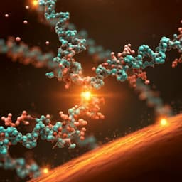
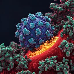
This groundbreaking research, conducted by Yusuke Higuchi and colleagues, presents an engineered high-affinity human ACE2 receptor that neutralizes SARS-CoV-2 effectively, showcasing its potential to combat the virus and prevent escape mutants. The study demonstrates significant protection against the virus in hamsters, suggesting a promising therapeutic approach.... show more
Introduction

The study addresses the urgent need for therapeutics resilient to SARS-CoV-2 mutational escape. SARS-CoV-2 infects cells via the spike RBD binding to ACE2. Although neutralizing antibodies often target the RBD, the virus can rapidly acquire escape mutations under antibody pressure. Soluble ACE2 (sACE2) acts as a decoy receptor with a theoretical advantage: mutations that reduce sACE2 binding should also reduce binding to native ACE2, compromising viral fitness. However, wild-type sACE2 binds RBD relatively weakly (KD ~20 nM), limiting therapeutic potential. The authors hypothesize that engineered ACE2 variants with greatly enhanced affinity for RBD can achieve potent neutralization comparable to monoclonal antibodies while resisting viral escape, and can provide therapeutic benefit in vivo.

Literature Review

Background literature indicates that coronaviruses, despite possessing proofreading, still accumulate mutations at rates that permit adaptation, including escape from monoclonal antibodies and convalescent plasma. The RBD is the major neutralizing epitope in convalescent sera. Prior work showed sACE2 can neutralize SARS-CoV-2 and is in clinical testing, but wild-type sACE2 has weaker affinity than clinical antibodies. Yeast-display engineering efforts previously identified higher-affinity ACE2 variants, including mutations in PD1 and PD2 regions that enhance RBD binding. Reports also documented rapid emergence of escape mutants against single antibodies and even cocktails or plasma in vitro. These findings motivate engineering ACE2 decoys with increased affinity and broad resilience to escape.

Methodology
  • Directed evolution in human 293T cells: The ACE2 protease domain was split into PD1 (residues 18–102) and PD2 (272–409) for independent mutagenesis via error-prone PCR (≈10 mutations/kb). Libraries (~10^5 variants) were expressed on 293T cell surfaces via lentiviral transduction at MOI <0.3. Cells were incubated with RBD fused to sfGFP. Flow cytometry quantified RBD binding (sfGFP) vs ACE2 expression (anti-HA Alexa 647). The top 0.05% of cells (high binding per expression, with surface ACE2 preserved) were sorted. Recovered ACE2 sequences were re-mutagenized for the next cycle. Three cycles were conducted for PD1; PD2 mutagenesis was also attempted post-PD1 selection.
  • Clone validation and competition assays: Selected ACE2 variants (e.g., 1-19, 2-51, 2-53, 3J38, 3N39) were produced as soluble ACE2-sfGFP (residues 18–615) to compete with cell-surface WT ACE2 for RBD binding in flow cytometry. Essential mutations for top clones (3N39, 3J113, 3J320) were identified by back-mutating nonessential changes, yielding minimal variants (v2 versions).
  • Binding kinetics: Surface plasmon resonance (Biacore T200) measured ACE2-His binding to captured RBD-Fc to derive KD and kinetic parameters. Mass photometry assessed stoichiometry of spike trimer binding with WT vs mutant ACE2.
  • Protein stability and formulation: Thermal shift assays (differential scanning fluorimetry) measured Tm. To extend half-life, ACE2 variants were produced as human IgG1 Fc fusions (ACE2-Fc). A designed disulfide (S128C/V343C) locked ACE2 in a closed conformation to abolish catalytic activity and enhance stability.
  • Neutralization assays: Pseudovirus neutralization in ACE2-expressing 293T cells measured IC50 values for SARS-CoV-2 and SARS-CoV-1 spikes. Authentic SARS-CoV-2 neutralization was tested in VeroE6/TMPRSS2 cells with qRT-PCR readouts of viral RNA and plaque assays where indicated.
  • Structural biology: The 3N39 ACE2-RBD complex was crystallized and solved at 3.2 Å (PDB 7DMU) to elucidate structural bases of affinity gains.
  • Escape mutation passage: Authentic SARS-CoV-2 was passaged serially (0.1 MOI) in VeroE6/TMPRSS2 cells under partially neutralizing concentrations of ACE2-Fc variants or a monoclonal antibody (H4). At each passage, 3×10^5 copies from partially neutralized wells were transferred. Viral RNA in supernatants was quantified; resistance emergence was assessed and escape mutations sequenced.
  • Variant testing: Neutralization of B.1.1.7 (UK), B.1.351 (South Africa), and P.1 (Brazil) variants was evaluated using pseudovirus and authentic isolates.
  • In vivo efficacy: Syrian hamsters received intranasal SARS-CoV-2 challenge (1.0×10^6 PFU). 3N39v2-Fc (20 mg/kg) or control-Fc was administered intraperitoneally 2 h post-infection (and in a separate cohort, at 2 dpi). Outcomes included body weight, lung micro-CT, lung viral titers (PFU, RNA copies), histopathology (H&E, antigen staining), and cytokine mRNA expression.
  • Pharmacokinetics: Single-dose i.p. ACE2-Fc in mice with plasma and lung concentrations measured by ELISA; half-life estimated using a two-phase assumption and one-phase modeling on day 1–7 data.
Key Findings
  • High-affinity ACE2 variants: After three PD1-directed evolution cycles, top mutants 3N39, 3J113, and 3J320 were identified. Minimal variants retained essential substitutions: 3N39v2 (A25V, K31N, E35K, L79F), 3J113v2 (K31M, E35K, Q60R, L79F), 3J320v2 (T20I, H34A, T92Q, Q101H).
  • Binding affinity (SPR): WT ACE2 KD = 17.63 nM. Mutants achieved KD values of 0.29 nM (3N39), 0.64 nM (3N39v2), 1.14 nM (3J113v2), and 3.98 nM (3J320v2), reflecting up to ~100-fold improvement with markedly slower off-rates.
  • Stoichiometry: Mass photometry showed mutant ACE2s (3N39, 3N39v2) predominantly form 3:1 ACE2:spike trimer complexes at slight excess, indicating all three RBDs on spike can be saturated when affinity is high.
  • Neutralization potency: Pseudotyped SARS-CoV-2 IC50 (µg/mL): WT 24.8; 3N39 0.056; 3N39v2 0.082; 3J113v2 0.33; 3J320v2 0.068. SARS-CoV-1 IC50 (µg/mL): WT 6.8; 3N39 0.011; 3N39v2 0.027; 3J113v2 5.3; 3J320v2 0.022. Authentic SARS-CoV-2 neutralization showed mutants effective at ~100-fold lower concentration than WT.
  • Structural basis (PDB 7DMU): Affinity gains arise from: (i) K31N/E35K disrupts an intramolecular salt bridge, enabling K35 to form a direct intermolecular H-bond with RBD Q493 and a ~1 Å approach of ACE2 α1-helix; (ii) L79F and A25V expand hydrophobic packing against RBD F486 and fill a back pocket, improving interaction energy (~2.76 kcal/mol gain estimated). 3N39 adopts a closed conformation similar to inhibitor-bound WT, yet closed conformation alone does not increase RBD affinity.
  • Disulfide-locked closed ACE2 (S128C/V343C): Abolished catalytic activity, did not alter RBD-binding affinity or competition, but markedly increased stability (Tm shifts: +12.5 °C WT, +7.0 °C 3N39, +11.5 °C 3N39v2).
  • Escape resistance: Under ACE2-Fc pressure, no escape mutants emerged over 15 passages, and passaged viruses remained sensitive. In contrast, under monoclonal antibody H4, resistance appeared by passage 4 with escape mutation F490V. Engineered ACE2-Fcs neutralized H4-resistant F490V and convalescent plasma escape mutants (A69-70/D796H; an N-linked glycan insertion variant).
  • Variant neutralization: Engineered ACE2-Fcs retained potent neutralization against B.1.1.7, B.1.351, and P.1 variants in pseudovirus assays; 3N39v2-Fc also neutralized authentic B.1.1.7 and P.1 alongside a reference antibody (REGN10933) comparison.
  • In vivo efficacy (hamsters): Therapeutic 3N39v2-Fc given 2 h post-challenge prevented weight loss (treated +7.3% vs control-Fc −4.3% at 5 dpi), reduced lung viral PFU and RNA copies, limited CT lung abnormalities, ameliorated histopathology, and decreased inflammatory/chemotactic cytokines (e.g., IL-6, IFN-γ, CXCL10, CCL3, CCL5, IL-10, TGF-β). Dosing at 2 dpi still reduced viral RNA and cytokines modestly but significantly.
  • Pharmacokinetics: Fc fusion extended plasma half-life to ~25–30 h (vs ~3 h reported for rsACE2), with measurable lung distribution after i.p. administration.
  • Safety/functional considerations: ACE2-Fc retains catalytic activity like WT; disulfide-locked closed variants eliminate enzymatic activity while preserving neutralization and stability, potentially reducing off-target vasodilatory effects.
Discussion

The study demonstrates that human cell-based directed evolution can yield ACE2 decoy receptors with ~100-fold higher affinity for SARS-CoV-2 RBD, translating to monoclonal antibody-like neutralization potency and robust activity against diverse variants. Structural analysis explains the affinity gains through optimized hydrogen bonding and hydrophobic packing, while stabilization via a disulfide-locked closed conformation enhances biophysical properties without compromising binding. Crucially, ACE2 decoys imposed minimal evolutionary escape in vitro across 15 passages, in stark contrast to rapid antibody escape, supporting the hypothesis that maintaining viral fitness constraints at the receptor interface curbs resistance. The work highlights advantages of mammalian display (native post-translational modifications) over yeast display and notes context dependence of mutations that limits simple combination strategies, suggesting orthogonal selection methods and additional structural insights may be needed for further improvements. The in vivo hamster data confirm therapeutic benefit with reduced viral burden and lung pathology. Pharmacokinetic extension via Fc fusion supports systemic utility, and Fc engineering choices (effector functions vs half-life) may be tailored to therapeutic vs prophylactic contexts.

Conclusion

Engineered ACE2 decoy receptors generated by mammalian cell-based directed evolution achieve sub-nanomolar RBD affinity, potent neutralization of SARS-CoV-2 (including major variants), and strong resistance to escape, with demonstrated therapeutic efficacy in a hamster model. Structural elucidation informs the design principles underlying affinity enhancement, and a disulfide-locked closed ACE2 offers improved stability and abrogated enzymatic activity for safety. These findings position engineered ACE2-Fc as a promising therapeutic strategy for COVID-19 and potentially future coronavirus threats. Future work should focus on minimizing immunogenicity, optimizing Fc for desired effector/half-life profiles, exploring orthogonal selection schemes to surpass current affinity ceilings, comprehensive safety profiling, dose optimization, and progression to clinical trials.

Limitations
  • The selection approach may have reached a practical limit as isolated variants already exhibit very slow dissociation; further affinity gains may not increase selection signals in FACS. Orthogonal methods may be necessary to surpass current ceilings.
  • Mutational effects are highly context-dependent; combinations with PD2 mutations from other studies reduced affinity, limiting straightforward additive design.
  • In vivo efficacy was demonstrated in hamsters with intraperitoneal dosing; translation to humans requires clinical evaluation, optimized dosing, route, and safety assessment.
  • Immunogenicity of engineered ACE2 variants was not evaluated; anti-drug antibody responses could pose risks, including potential cross-reactivity to endogenous ACE2.
  • Although catalytic activity can be eliminated by disulfide locking, comprehensive assessment of hemodynamic or off-target effects was not performed.
  • Pharmacokinetic analyses were limited and model-dependent due to i.p. administration; human PK/PD may differ.
Listen, Learn & Level Up
Over 10,000 hours of research content in 25+ fields, available in 12+ languages.
No more digging through PDFs, just hit play and absorb the world's latest research in your language, on your time.
listen to research audio papers with researchbunny