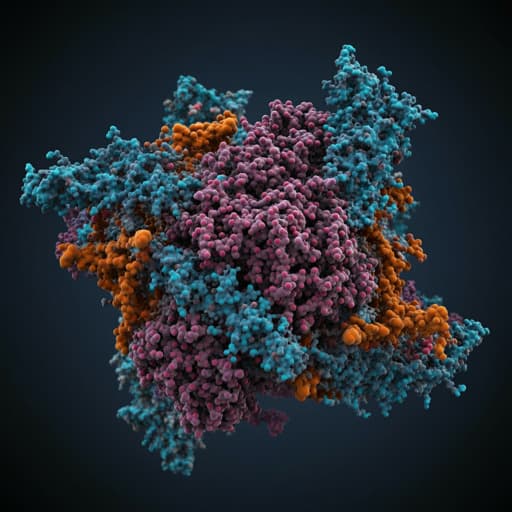
Does the SARS-CoV-2 Spike Receptor-Binding Domain Hamper the Amyloid Transformation of Alpha-Synuclein after All?
Y. Stroylova, A. Konstantinova, et al.
Explore these studies to deepen your understanding
Adjacent work that informs or extends this paper's methodology and findings.
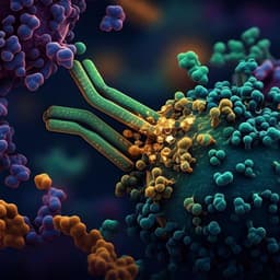
A therapeutic neutralizing antibody targeting receptor binding domain of SARS-CoV-2 spike protein
C. Kim, D. Ryu, et al.
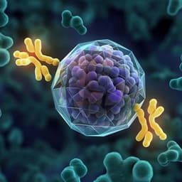
DNA origami presenting the receptor binding domain of SARS-CoV-2 elicit robust protective immune response
E. Oktay, F. Alem, et al.
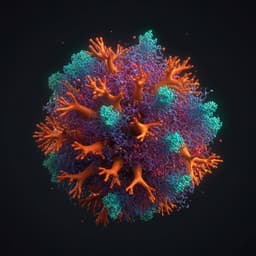
Analysis of the N-glycosylation profiles of the spike proteins from the Alpha, Beta, Gamma, and Delta variants of SARS-CoV-2
D. Wang, J. Baudys, et al.

Risk factors for and pregnancy outcomes after SARS-CoV-2 in pregnancy according to disease severity: A nationwide cohort study with validation of the SARS-CoV-2 diagnosis of Nordic Federation of Societies of Obstetrics and Gynecology (NFOG)
A. J. M. Aabakke, T. G. Petersen, et al.


