Chemistry
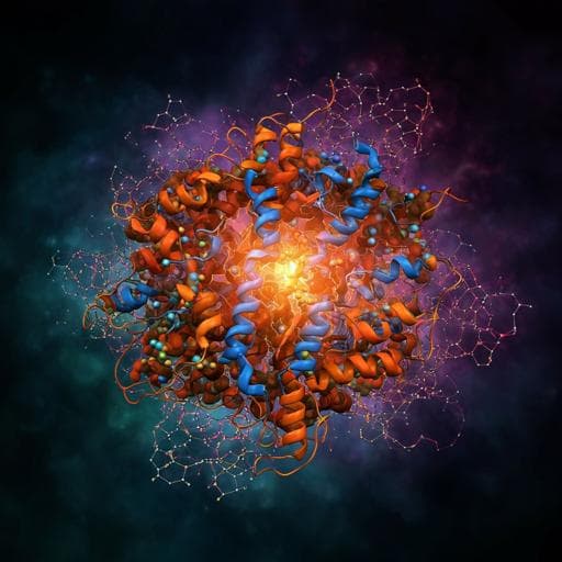
Directed ultrafast conformational changes accompany electron transfer in a photolyase as resolved by serial crystallography
A. Cellini, M. K. Shankar, et al.
Delve into the fascinating world of charge-transfer reactions in proteins with groundbreaking research from Andrea Cellini, Madan Kumar Shankar, and team. Their study reveals the intricate structural dynamics involved in electron transfer processes within the *Drosophila melanogaster* photolyase, highlighting the role of conserved tryptophans and other key residues, and challenging longstanding theories in the field.
~3 min • Beginner • English
Introduction
Electron transfer in proteins underpins key biological processes, including photosynthesis, respiration, oxidative stress defense, and DNA repair. Photolyases/cryptochromes provide a paradigm where a photoexcited FAD cofactor is reduced via a conserved chain of tryptophans, forming long-range radical pairs. Despite extensive spectroscopic work, how protein structural dynamics guide and stabilize charge transfer at ultrafast timescales remains unresolved, and the applicability of classical Marcus theory is questionable under non-ergodic protein dynamics. Prior estimates of free-energy and reorganization energies rely on assumptions that may not hold, and simulation predictions have not fully matched experiments. The study aims to directly resolve the site-specific, ultrafast structural responses that accompany electron transfer along the tryptophan tetrad in Drosophila melanogaster (6-4) photolyase, to clarify timing, directionality, and the role of the local environment in facilitating charge transport.
Literature Review
Previous studies established that in photolyases/cryptochromes, electron transfer proceeds from photoexcited FAD to a nearby tryptophan, then along a conserved tryptophan chain, generating a long-range radical pair (Aubert et al., 2000; Liu et al., 2013). Photolyases then undergo a second photoreduction to yield FADH− for DNA repair, with proton transfer events on millisecond timescales. Marcus theory has framed electron transfer in terms of free-energy gains and reorganization energies, but proteins exhibit multi-timescale relaxations potentially invalidating linear solvent response assumptions and leading to non-ergodic behavior (Sumi & Marcus, 1986; Lu et al., 2020). Reported per-step free-energy gains are a few hundred meV with moderate reorganization energies (<1 eV), yet extracting individual tryptophan-to-tryptophan rates spectroscopically is difficult. Molecular simulations have predicted energetics for the (6-4) photolyase tryptophan tetrad, but even recent self-consistent treatments showed discrepancies with experimental kinetics. Time-dependent Stokes shift studies suggested water responds within a few picoseconds, combined protein–water motions in tens of picoseconds, and larger protein breathing on hundreds of picoseconds, but these lack structural specificity. Time-resolved serial crystallography (SX) has resolved ultrafast structural changes in other proteins and, at longer times, in photolyases. The present work fills the gap by capturing femto- to picosecond structural snapshots during the first photoreduction and subsequent electron hopping steps.
Methodology
The authors employed femtosecond time-resolved serial crystallography (SX) at SwissFEL (Alvra instrument) on microcrystals of Drosophila melanogaster (6-4) photolyase embedded in 22% hydroxyethyl cellulose. Photoexcitation: 150 fs pulses at 474 ± 12.5 nm, 9.6 µJ per pulse, focused to 80 × 86.4 µm² (1/e²), fluence 1.4 mJ mm−2, within the one-photon regime as confirmed by fluence-dependent DED scaling. X-ray probe: 12.06 keV, 50 fs (r.m.s.d.), 100 Hz, detected by JUNGFRAU 4M. Time points: dark, 400 fs, 1 ps, 2 ps, 20 ps, 300 ps, and 100 µs. Data volumes: e.g., 42,547 indexable frames (dark), 21,576 (400 fs), 98,625 (20 ps), etc. Crystallographic processing used CrystFEL (xgandalf indexing; peakfinder8), Ambigator for indexing ambiguity resolution, and Partialator for scaling/post-refinement (xsphere partiality model). Difference electron density (DED) maps were computed as ΔF = w(F(light) − F(dark)) with dark phases; w down-weights low-resolution amplitudes with large errors. Maps used 16–1.9 Å resolution cutoffs, with light/dark scaling via ccp4 scalit. Dark structures were refined in PHENIX (molecular replacement using 3CVY; manual model building in Coot). Photoinduced changes were modeled by real-space refinement against extrapolated maps Fc = Fc(dark) + NΔFc with extrapolation factor N ≈ 14 estimated from negative DED analysis. Model validation included comparing observed vs calculated ΔFc maps and real-space Pearson correlation. Region-specific DED integration quantified kinetics around predefined residue clusters proximal to FAD/Trp407 (region 1), Trp384 (region 2), and Trp381 (region 3). Complementary femtosecond transient absorption spectroscopy used a Ti:sapphire amplifier (800 nm, 3 kHz). Pump at 470 nm (TOPAS), white-light continuum probe (CaF2), with <3 ns delay range; pump fluence set ~30% below typical literature levels to ensure linear regime. Samples were temperature-controlled (~5 °C) and translated to avoid photodamage. Data were dispersion-corrected and globally fitted (sum of exponentials convolved with IRF) using TIMP/Glotaran to extract evolution-associated decay spectra and time constants. Protein expression/purification followed established protocols with Heparin affinity and size-exclusion chromatography; photoactivity validated by UV–Vis before crystallization; microcrystals grown via seeding and embedded in carrier medium using gentle syringe mixing to preserve crystallinity.
Key Findings
- Time-resolved SX reveals significant DED features tracking electron flow from FAD to tryptophans: region 1 (near FAD/Trp407) peaks at 1 ps; region 2 (around Trp384) peaks at 20 ps; region 3 (around Trp381) rises at 300 ps and 100 µs, indicating charge arrival at the terminal tryptophan.
- Transient absorption spectroscopy shows charge separation between FAD and Trp407 within <520 fs, inter-tryptophan transfer time constants of ~12 ps and ~450 ps, and long-lived charges beyond 1 ns.
- Around FAD N5 and Trp407, Asn403 exhibits immediate and evolving structural response: its side chain twists, moving the carbonyl away from FAD N5 from 400 fs to 20 ps. Distances (Å) from Table 1: Asn403(C=O)–FAD(N5) changes from dark 3.66 to 4.59 (400 fs), 3.90 (1 ps), 4.21 (2 ps), 3.87 (20 ps), 4.06 (300 ps). Asn403–Trp407 remains essentially unchanged (~2.9–3.1 Å), indicating a rigid Trp407 environment.
- Water dynamics are time-delayed and spatially evolving near FAD N5: transient positive DED features appear at 2 ps (features IV/V) and more pronounced at 20–300 ps (feature VI), consistent with water molecules hydrogen bonding to and stabilizing the reduced FAD. No significant water features at 400 fs–1 ps; the water response lags side-chain motions.
- The conserved Asp397–Arg368 salt bridge on the opposite side of FAD responds ultrafast: correlated DED (feature VII) from 400 fs, maximal at 1–2 ps, decaying by 300 ps. Light-state refinements show a transient increase in the salt-bridge distance due to Asp397 rotation: e.g., Asp397(OD1)–Arg368(NH1) changes from 2.98 Å (dark) to 3.33 Å (1 ps), then relaxes toward 3.06 Å (300 ps); OD2–NH2 from 2.75 Å (dark) to 2.67–2.84 Å.
- On FAD, no clear isoalloxazine ring bending is detected; negative features on the pyrophosphate appear at 1–2 ps (uncertain interpretation). Evidence for a side reaction consistent with transient adenine oxidation appears at 2–300 ps (feature IX), with disruption of the adenine–Wat70 hydrogen bond.
- Along the tryptophan tetrad, no strong DED on tryptophan side chains is observed above 3.5σ except at Trp381 from 20 ps onward. The absence of structural changes near Trp407 despite sub-ps electron transfer suggests a rigid local environment or rapid onward transfer to Trp384.
- A strong, delayed positive DED feature adjacent to the sulfur of conserved Met408 (4.1 Å from Trp384) appears from 1–20 ps (max at 2 ps) and decays by 300 ps, consistent with transient oxidation of Met408 mediated by a nearby water molecule, implicating Met408 as an active participant in the redox chain.
- At the terminal Trp381, pronounced DED at 100 µs (with signals already at 20 and 300 ps) indicates arrival of charge and structural rearrangements: breaking of the Pro329–Trp381 backbone hydrogen bond in the dark state, Trp381 side-chain movement, and Asn490 approaching Trp381. The 100 µs state is assigned to TrpH•+ prior to deprotonation.
- Overall, the protein exhibits highly directed and timed conformational responses—Asn403 twisting, ultrafast salt-bridge modulation, water reorganization, transient Met oxidation—synchronized with electron hopping, challenging linear solvent response assumptions.
- Fluence dependence confirms one-photon regime: lowering effective absorbed photons by ~1.8 reduces DED amplitude; further lowering eliminates signal.
Discussion
The study directly visualizes ultrafast, site-specific structural dynamics that accompany and likely facilitate electron transfer in a (6-4) photolyase. The immediate response of the Asp397–Arg368 salt bridge and rapid Asn403 side-chain twist upon FAD photoreduction reflect targeted electrostatic re-organization around the chromophore, while picosecond-delayed water relocation completes stabilization of the reduced FAD. These temporally staggered but spatially coordinated adaptations provide a mechanistic basis for achieving swift charge separation with minimal recombination and for steering electrons along chemically identical tryptophans via environmental tuning. The transient involvement of Met408 near Trp384 suggests that conserved peripheral residues and associated waters modulate redox potentials and coupling within the chain, adding an active role for methionine oxidation states in guiding electron flow. The observed kinetics (sub-ps FAD←Trp407 transfer, picosecond-scale Trp-to-Trp steps, and arrival at Trp381 within hundreds of picoseconds) agree qualitatively with spectroscopy and crystallography, but the nonuniform, directed structural responses question the validity of treating the environment with a linear solvent response within classical Marcus theory. Instead, the results support non-ergodic protein dynamics and evolutionarily tuned, fast fluctuations as critical determinants of charge-transfer efficiency in photolyases/cryptochromes.
Conclusion
This work captures a molecular movie of ultrafast, directed conformational changes that accompany electron transfer in Drosophila (6-4) photolyase. Key contributions include: (1) resolving femto-to-picosecond structural responses at residues Asn403 and the Asp397–Arg368 salt bridge that stabilize FAD reduction; (2) revealing delayed, dynamic water reorganization near FAD N5 that completes charge stabilization; (3) identifying conserved Met408 near Trp384 as an active participant, likely via transient oxidation; and (4) observing structural signatures of charge arrival at Trp381 within hundreds of picoseconds and persistent changes at 100 µs. Together, these results demonstrate orchestrated, residue- and water-specific motions that guide electron hopping and challenge linear-response assumptions underlying Marcus theory in proteins. Future work could extend time-resolved crystallography across broader time windows and to other photolyases/cryptochromes, dissect mutational effects at key residues (Asn403, Asp397/Arg368, Met408), and integrate improved non-equilibrium theoretical models to quantitatively link structure, dynamics, and electron-transfer energetics.
Limitations
- DED signals are small and localized; most tryptophan side chains (except Trp381 at later times) show no DED above 3.5σ, limiting direct visualization of radical localization on indoles.
- FAD conformational changes (e.g., isoalloxazine bending) are not clearly resolved; pyrophosphate features at 1–2 ps are difficult to interpret.
- The photolyase crystals undergo FAD reoxidation, potentially faster than in other systems, complicating comparisons to microsecond dynamics reported for different photolyases.
- Spectroscopy did not resolve the Trp3→Trp4 transfer explicitly; assignment relies on crystallographic timing and prior expectations.
- Modeling of transient waters and side-chain rotamers carries uncertainty; alternative Asn403 models failed to match observed DED but underscore sensitivity to model assumptions.
- Present-address affiliations for some authors indicate potential differences between experimental and current institutional settings; not a scientific limitation but relevant for attribution.
Related Publications
Explore these studies to deepen your understanding of the subject.

