Food Science and Technology
Decellularised plant scaffolds facilitate porcine skeletal muscle tissue engineering for cultivated meat biomanufacturing
P. Murugan, W. S. Yap, et al.
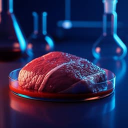
Cultivated meat aims to address sustainability, safety, environmental, and animal welfare issues by growing muscle tissues in controlled conditions using tissue engineering. Structured products require suitable cells, media, scalable bioprocesses, and especially edible scaffolds that provide macro-porosity, mechanical support, and anisotropic architecture to align myotubes for realistic texture. Plant-derived, decellularised tissues offer sustainable, abundant, animal-free scaffolds that preserve inherent vascular architectures while removing cellular components. Prior plant scaffolds (e.g., spinach leaves, grasses, broccoli florets) supported cell growth but were limited by thickness or lacked clear myogenic differentiation outcomes. This study targets that gap by developing an edible decellularised plant scaffold from asparagus with aligned vascular bundles to promote cell attachment and alignment, supporting proliferation and differentiation of C2C12 myoblasts and porcine adipose-derived mesenchymal stem cells (pADMSCs). The aim is to create a co-cultured muscle–fat cultivated meat prototype and evaluate scaffold biocompatibility, myogenesis, gene/protein expression, cooking durability, and texture relative to pork loin.
The authors review scaffold requirements for structured cultivated meat, emphasizing macro-porosity, anisotropy, and edible/biodegradable compositions with favourable organoleptic profiles. Plant decellularisation yields scaffolds with preserved 3D architectures that can reduce reliance on animal agriculture. Prior work includes decellularised spinach and grass supporting bovine satellite cells and C2C12 with vascular networks but limited thickness requiring stacking. Decellularised broccoli florets functioned as microcarriers for bovine cells in dynamic culture without demonstrating muscle differentiation. Previous asparagus-based scaffolds for skeletal muscle used non-edible fibronectin for biofunctionalisation. Collectively, these studies suggest plant-derived scaffolds are promising yet require improved thickness, alignment, edibility, and demonstration of long-term proliferation and myogenesis, as well as inclusion of adipose components to better mimic conventional meat.
Scaffold preparation: White asparagus (Asparagus officinalis) stems were cut longitudinally along vascular bundles and punched to 12 mm diameter discs. Decellularisation used 1% (w/v) SDS for 88 h (solution refreshed after 24 h), followed by 1% (v/v) Triton X-100 for 24 h, 100 mM CaCl2 for 24 h, 70% (v/v) ethanol for 30 min, triple DI water rinses, an additional 24 h in 100 mM CaCl2, rinsing to remove surfactant residue, freezing, and lyophilisation (≥48 h). Scaffolds were stored at −20 °C. Characterisation: DNA content was quantified (CYQUANT assay) on cryomilled raw and decellularised samples. FTIR identified scaffold composition; EDX provided elemental composition. Mechanical testing determined Young’s modulus. SEM imaged surface topography. Micro-CT (SkyScan 1272) assessed 3D architecture, pore size distribution, porosity and connectivity with CTAn analysis. Cell sourcing and culture: C2C12 myoblasts (ATCC CRL-1772) were cultured in DMEM +10% FBS +1% Pen/Strep at 37 °C, 5% CO2 (passages 4–6). Porcine adipose-derived MSCs (pADMSCs) were isolated from subcutaneous adipose tissue via collagenase II digestion, filtration, RBC lysis, expansion in DMEM +10% FBS +P/S (passages 4–6). Multipotency was verified by adipogenic, osteogenic, and chondrogenic staining in 2D. Scaffold biofunctionalisation and 3D myogenesis: Scaffolds were sterilised (70% ethanol), PBS-washed, and biofunctionalised over 3 days. They were pre-incubated in proliferation medium (PM). Static seeding was performed on both sides: C2C12 (100,000 cells per side; 6 h between sides) cultured 7 days in PM (DMEM +10% FBS +1% Pen/Strep) to confluency, then switched to differentiation medium (DMEM +2% horse serum +1% Pen/Strep) for 9 days. For pADMSCs, the same seeding approach with 12 h incubation per side, PM (DMEM +20% FBS +1% Pen/Strep) for 7 days to confluency, then DM for 17 days. Adipogenesis and co-culture: pADMSCs were differentiated to adipocytes using a proprietary scaffold-based approach with chemical cues over 7 days (LipidTOX Red used for verification). On Day 17 of pADMSC myogenesis in the scaffold, 1×10^6 pADMSC-derived adipocytes were seeded on top; co-culture maintained under serum-reduced conditions for 5 days with alternate-day media changes. Assays and imaging: Viability and coverage were assessed by live/dead/nuclear staining (calcein AM, ethidium homodimer-1, Hoechst) on PM Days 2 and 7 and DM Day 7; metabolic activity was quantified via PrestoBlue. Myogenic differentiation was evaluated by immunofluorescence: C2C12 (MHC, MYOG, F-actin), pADMSCs (MHC, DES, F-actin), with confocal imaging and EDF/3D rendering. Cell alignment was quantified from phalloidin images (ImageJ Orientation plugin; CircStat Kappa). CK activity was measured (Sigma MAK116) and normalised to total protein (BCA) across proliferation and differentiation time points. qPCR quantified MYH1 and MYOG expression (TaqMan; normalised to Rn18S; 2^−ΔCt). SEM/FESEM examined morphology (CPD dried, Pt-sputter coated). Co-cultures were stained with muscle markers plus LipidTOX for adipocytes. Micro-CT quantified pore metrics. Thermal stability was evaluated by TGA (Supplementary). Cooking and texture: Scaffolds (with/without cells) and pork loin were cut to identical dimensions, pan-fried in olive oil (80 °C, 600 W, 30 s per side) and cooled. Texture profile analysis (TA.XTPlusC; 75 mm platen; 50% deformation; 2 mm/s; two-cycle; 5 s interval; n=5) assessed hardness, springiness, cohesiveness, chewiness in raw and cooked states. Statistics used Brown-Forsythe and Welch ANOVA for viability; one-way ANOVA with Tukey’s post hoc for CK, qPCR, TPA; data reported as mean ± s.d.
- Decellularisation efficacy: DNA content reduced from 978 ± 62 ng/mg (raw) to 254 ± 60 ng/mg (DPS) (n=2; P<0.01).
- Composition and mechanics: FTIR indicated cellulose, hemicellulose, pectin; Young’s modulus 4.9 ± 1.12 kPa (n=5; ns vs raw), within range between adipogenic (~3 kPa) and myogenic (10–18 kPa) benchmarks.
- Architecture: DPS discs averaged 10.1 ± 0.6 mm diameter and 2.0 ± 0.5 mm thickness (n=5). Pore diameters 8–80 µm with a mode at 44 µm (10.5% of volume). Micro-CT showed total porosity ~93.5% and open pore connectivity 93.55%; closed porosity 0.0015%; open surface area 99.99%.
- C2C12 proliferation and myogenesis: Viability increased 2.94-fold from Day 2 to Day 7 in PM (n=5; P<0.01 to <0.001). Cells aligned along vascular bundles; SEM showed aligned myotubes. Immunofluorescence showed MHC and MYOG expression emerging by DM-Day 2, stronger by DM-Days 5–9, with multinucleated myotubes. CK activity increased up to 10.92-fold at DM-Day 9 vs PM-Day 6 (n=3; P<0.05). qPCR: On DM-Day 7, MYH1 up 16.7-fold vs Day 0 and 2.98-fold vs PM-Day 6; MYOG up 5.3-fold vs Day 0 and 2.78-fold vs PM-Day 6 (n=3; P<0.01 to <0.0001).
- pADMSC proliferation and myogenesis: Viability increased 3.64-fold from Day 2 to Day 7 in PM (n=5; P<0.01 to <0.001). Immunostaining showed MHC and DES expression with elongated multinucleated myotubes; SEM confirmed alignment along bundles. CK activity rose to 1.95-fold at DM-Day 14 vs PM-Day 7 (n=3; ns).
- Co-culture: pADMSC-derived adipocytes adhered atop differentiated muscle within DPS. LipidTOX confirmed intracellular neutral lipid accumulation; muscle markers (MHC, DES) verified myotubes. SEM displayed round adipocytes on elongated myotubes, maintaining phenotypes through Day 5 of co-culture.
- Texture and cooking: TGA indicated thermal stability of DPS. TPA on raw samples showed no significant differences in hardness, springiness, chewiness among scaffold variants vs pork loin; cohesiveness differed significantly. After pan-frying, CM prototypes differed significantly from pork loin in hardness and chewiness (n=5; P<0.05 to <0.0001). Visible browning occurred upon cooking.
The study demonstrates that decellularised asparagus-derived plant scaffolds (DPS) provide an edible, aligned, macro-porous platform that supports attachment, alignment, proliferation, and myogenic differentiation of both C2C12 myoblasts and primary porcine adipose-derived MSCs. The scaffold’s vascular bundle alignment promoted myotube organization, while its porosity facilitated nutrient and gas exchange. Despite DNA content exceeding regenerative medicine decellularisation thresholds, prior plant-scaffold studies and the edible nature of asparagus render this non-critical for cultivated meat. The robust C2C12 differentiation (protein markers, elevated CK, and upregulated MYH1 and MYOG) and successful pADMSC myotube formation confirm DPS suitability for muscle tissue engineering. Importantly, the co-culture of pADMSC-derived muscle and fat cells on a single, edible scaffold produced a hybrid tissue more representative of conventional meat structure, advancing biological and sensory mimicry. Textural analyses suggest raw prototypes approximate pork loin on several parameters, whereas cooking introduces significant differences in hardness and chewiness, informing further optimisation of scaffold composition, cell density/ratio, and processing to approach conventional meat mouthfeel. Compared with prior plant scaffolds that were thin or required non-edible biofunctionalisation, this approach delivers an edible, mechanically stable scaffold with improved structural features, offering a pathway toward scalable, structured cultivated meat products.
This work establishes decellularised asparagus as a promising edible scaffold for cultivated meat, enabling aligned, volumetric skeletal muscle tissue formation and supporting the co-culture of muscle and adipose cells to better mimic conventional meat. DPS supports cell viability, alignment, and myogenesis with clear protein and gene expression and increased CK activity. The prototype exhibits comparable raw textural attributes to pork loin on several metrics, with differences emerging after cooking. These findings provide a foundation for developing structured, scalable cultivated meat products using plant-derived scaffolds. Future work should streamline decellularisation with food-safe surfactants, integrate perfusion/bioreactor strategies leveraging scaffold porosity for scale-up, tune scaffold mechanics and composition for cooked texture convergence, optimise muscle–fat ratios and maturation, and expand DPS to other edible plant species and geometries for diverse product formats.
- Residual DNA content after decellularisation (~254 ± 60 ng/mg) exceeds conventional regenerative medicine thresholds, though not critical for edible applications.
- Decellularisation/purification was lengthy and required surfactants; future use of safer, food-grade surfactants and industrial processes is needed.
- Media perfusion and scale-up through the scaffold’s porous network were not investigated.
- Texture differences persisted after cooking (hardness and chewiness), indicating further optimisation is required.
- Some assays had small sample sizes (e.g., DNA quantification n=2) and CK increase in pADMSCs was not statistically significant, warranting larger studies.
- Mechanical tests were performed on dry scaffolds, while TPA involved hydrated/cooked conditions, potentially introducing condition-dependent variability.
Related Publications
Explore these studies to deepen your understanding of the subject.