Medicine and Health
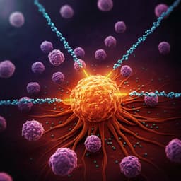
Chemotaxis-driven delivery of nano-pathogenoids for complete eradication of tumors post-phototherapy
M. Li, S. Li, et al.
This groundbreaking research by Min Li and colleagues unveils a revolutionary nano-pathogenoid system that transcends biological barriers in drug delivery, significantly boosting the power of photothermal therapy against tumors. Their innovative approach offers complete tumor eradication in mice with a combination of therapies, paving the way for advanced cancer treatments.
~3 min • Beginner • English
Introduction
Cancer initiation and therapy often involve inflammatory responses that recruit neutrophils capable of chemotaxis, extravasation, and deep tissue penetration. Conventional synthetic nanoparticles lack chemotactic behavior and face major biological barriers including rapid clearance by the mononuclear phagocyte system, poor extravasation, and limited interstitial penetration. Prior neutrophil-based delivery strategies either load nanoparticles ex vivo (limited by short neutrophil lifespan and logistics) or aim to hitchhike circulating neutrophils in situ via specific ligands, but more generalizable and efficient approaches are needed. The authors propose pathogen-mimicking nano-pathogenoids (NPNs): PEG-b-PLGA nanoparticles cloaked with bacterial outer membrane vesicles (OMVs) to present pathogen-associated molecular patterns (PAMPs) that are recognized by neutrophil pattern recognition receptors (TLR2/TLR4), enabling in situ capture by circulating neutrophils. Photothermal therapy (PTT) is used to induce acute intratumoral inflammation and chemokine gradients (e.g., CXCL1, MIP-2) that attract neutrophils, thereby enhancing delivery of neutrophil-laden NPNs to residual microtumors. The study aims to demonstrate that this chemotaxis-driven, neutrophil-hitchhiking delivery improves tumor targeting, overcomes vascular and interstitial barriers, promotes release of NPNs via NETosis within inflamed tumors, and eradicates tumors when combined with cisplatin after PTT.
Literature Review
The study builds on evidence that inflammation recruits neutrophils which exhibit robust chemotaxis and tissue infiltration. Conventional nanoparticles are limited by opsonization and clearance (MPS), vascular barriers, and dense tumor interstitium. Cell-mediated delivery has been explored, including neutrophils as carriers. Ex vivo loading of neutrophils faces viability and practicality constraints. In situ hitchhiking efforts using denatured albumin nanoparticles or anti-CD11b-decorated particles have shown feasibility but may lack generalizability. OMVs from Gram-negative bacteria carry PAMPs (e.g., LPS, lipoproteins) that engage neutrophil TLR4/TLR2, mimicking pathogens without the replicative risks of live bacteria. Phototherapies (PTT/PDT) can create inflammatory microenvironments but are limited by light penetration leading to residual disease and recurrence; combining PTT with targeted chemotherapy has been proposed to improve outcomes. This work integrates these strands by using OMV-coated nanoparticles to target neutrophils in circulation and PTT to prime tumors for chemotaxis-driven delivery.
Methodology
Materials and nanoparticle preparation: PEG-b-PLGA was synthesized by ring-opening polymerization. A near-infrared photothermal polymer (PBIBDF-BT) was synthesized and encapsulated into PEG-b-PLGA micelles to form NPs@PBT for PTT. Gold nanorods (GNRs) were also synthesized for control PTT studies. A hydrophobic Pt(IV) cisplatin prodrug was synthesized and encapsulated into PEG-b-PLGA to form NPs@Pt. Fluorescent dyes DiO/DiD were used for labeling. OMVs were prepared from Escherichia coli culture supernatants by filtration, ultrafiltration, and ultracentrifugation. Nano-pathogenoids (NPNs) were produced by extruding PEG-b-PLGA nanoparticles with OMVs (w/w 5:1 NP:OMV), yielding core–shell particles with OMV coatings.
Characterization: Dynamic light scattering (size, zeta potential), TEM imaging, and serum stability assays were performed. NPs@PBT photothermal performance under an 808 nm laser was calibrated using a square-wave-controlled duty cycle to maintain desired temperatures.
Cell lines and animal models: EMT6 murine breast carcinoma and CT26 colon carcinoma cells were used in BALB/c mice. EMT6-EGFP cells enabled tumor cell tracking. Tlr4−/− mice and TLR2 blockade were used to probe receptor involvement.
PTT and inflammation priming: Mice received i.v. NPs@PBT (or GNRs). After 12 h, tumors were irradiated with an 808 nm laser and temperature maintained at 40–45 °C for 2–10 min via duty-cycle control. Tumor/blood were harvested at defined time points for flow cytometry and ELISA (CXCL1, MIP-2). A CXCR2 inhibitor (SB225002) tested chemokine-receptor dependence.
Neutrophil recruitment assays: Flow cytometry quantified CD11b+Ly6G+ neutrophils in tumors and blood over time after PTT and versus tumor size, temperature, and duration. Adoptive transfer of DiD-labeled bone marrow neutrophils assessed tumor homing by in vivo imaging (IVIS), ex vivo organ imaging, and flow cytometry.
Intravital microscopy: Dorsal skinfold window chamber EMT6 tumors were imaged after PTT; PE-anti-Ly6G labeled neutrophils and DiO-labeled vessel tracers visualized intravascular rolling/adhesion, capillary crawling, extravasation, and interstitial migration. Tumor sections were stained for Ly6G and CD31 to quantify intravascular vs stromal neutrophils.
Neutrophil hitchhiking by NPNs: After i.v. DiO-labeled NPs or NPNs into PTT-treated mice, flow cytometry quantified the percentage and GMFI of NPN+ neutrophils in blood and tumors, and distribution among immune subsets. Tlr4 knockout and anti-TLR2 antibody tested receptor roles. Serum cytokines (IL-6, TNF-α) assessed transient systemic inflammation.
Tumor accumulation: IVIS quantified DiD-NPN accumulation in tumors with/without PTT; ex vivo organ imaging assessed biodistribution.
Cargo release and intercellular transfer: In vitro, PMA (100 nM) induced NETosis in neutrophils loaded with NPs to evaluate release and subsequent uptake by EMT6 cells (confocal microscopy) and cytotoxicity of released NPNs@Pt. In vivo, intratumoral injection of DiD-neutrophils containing NPs@DiO into PTT-treated tumors quantified decrease in NP+ transferred neutrophils and increased NP uptake by tumor cells via flow cytometry and IF; NETosis marker Cit-H3 confirmed in vivo.
Therapeutic studies: Single-treatment study (ten groups) compared PBS, cisplatin, OMVs, NPs@Pt, NPNs@Pt, PTT alone, and combinations (PTT + cisplatin/OMVs/NPs@Pt/NPNs@Pt). PTT was 40–41 °C for 5 min; cisplatin 2 mg/kg. Tumor volumes were measured bi-daily; tumors were harvested for H&E, Ki67, and TUNEL staining. Validation: CT26 model with low neutrophil infiltration, neutrophil depletion (anti-Ly6G), and NETosis inhibition (PAD inhibitor BB-Cl-amidine) tested mechanism dependence. Two-treatment regimen: PTT on days 0 and 3 plus i.v. NPs@Pt or NPNs@Pt; tumor growth, tumor-free rates, body weight, and organ histology were assessed. Statistical analyses used unpaired two-tailed Student’s t-tests with significance at P<0.05.
Key Findings
- PTT induces acute tumor inflammation and neutrophil recruitment: In EMT6 tumors, CD11b+Ly6G+ neutrophils in tumors significantly increased within 48 h, peaking at 4 h post-PTT (40 °C, 5 min). Blood neutrophils rose within 12 h then declined by 96 h. Higher PTT temperature (42–45 °C) and longer irradiation (up to 10 min) increased infiltration. Recruitment decreased with larger tumors, likely due to reduced light penetration; phenomenon was tumor-type independent (EMT6, CT26). CXCL1 and MIP-2 levels rapidly increased post-PTT; CXCR2 inhibition reduced recruitment.
- Adoptively transferred neutrophils preferentially home to PTT-primed tumors: IVIS showed ~2.4-fold higher tumor fluorescence at 16–24 h and ~2.7-fold higher ex vivo tumor signal at 24 h in PTT-treated mice vs controls; flow cytometry showed ~4-fold more transferred neutrophils in tumors at 10 h.
- Intravital imaging confirms barrier traversal: After PTT, ~120 neutrophils/mm² of vessels vs ~21/mm² in controls; time-lapse imaging captured rolling/adhesion, capillary deformation/crawling, and extravasation within minutes. Overall neutrophil density in PTT-treated tumors was ~3.8× controls, with more cells in interstitial stroma.
- NPN design and hitchhiking: OMV coating changed zeta potential from −18.1±0.5 mV (NPs) to −4.16±0.32 mV (NPNs), similar to OMVs (−3.96±1.18 mV); diameter increased ~7 nm, consistent with lipid layer thickness; particles were serum-stable. In blood 4 h post-injection, ~41% of neutrophils contained NPNs vs ~10% with uncoated NPs; neutrophils captured ~83.4% of total circulating NPNs among major immune cells (vs monocytes 9.6%, T cells 2.6%, B cells 3.3%, NK 1.1%). In tumors, ~19% of neutrophils contained NPNs vs ~7% with NPs. TLR4 knockout markedly reduced neutrophil uptake; TLR2 blockade reduced targeting by ~25%, implicating TLR4>T̄LR2 in recognition. Serum IL-6 and TNF-α transiently increased at 4 h post-NPNs then normalized by 24 h.
- Enhanced tumor accumulation: IVIS showed significantly greater DiD-NPN accumulation in PTT-treated tumors vs controls; ex vivo imaging confirmed increased tumor uptake without significant changes in major organs. NPN targeting after PTT exceeded EPR-based PEG-b-PLGA NP accumulation.
- Cargo release and tumor cell uptake under inflammation: PMA induced NETs and release of cargo from neutrophils; EMT6 cells internalized ~4× more NPs under inflammatory vs normal conditions. In vivo, after PTT, the percentage of transferred neutrophils still containing NPs decreased by 77.6%, indicating release; more NPs were detected in tumor cells; NETosis (Cit-H3) was confirmed in tumors.
- Therapeutic efficacy: Single treatment: PTT + NPNs@Pt provided the strongest tumor suppression, with ~79% greater inhibition vs PTT + NPs@Pt and reported 60% tumor-free rate with ~97% tumor growth inhibition. Histology showed highest apoptosis (TUNEL) and lowest proliferation (Ki67). OMVs alone or cisplatin alone had modest effects; nanocarriers (NPs@Pt) outperformed free cisplatin. Mechanism validation: In CT26 tumors with low neutrophil infiltration, PTT + NPNs@Pt did not outperform PTT + NPs@Pt; neutrophil depletion (anti-Ly6G) or NETosis inhibition (BB-Cl-amidine) significantly reduced efficacy and survival, confirming dependence on neutrophil carriage and cargo release via NETosis.
- Two-treatment regimen: PTT + NPNs@Pt on days 0 and 3 completely eradicated EMT6 tumors in all treated mice (100% tumor-free), whereas PTT + NPs@Pt yielded ~71% tumor growth inhibition with 40% tumor-free. NPNs@Pt alone inhibited growth (~52% vs NPs@Pt) with 20% tumor-free. Transient OMV-associated weight loss resolved within 5 days; no obvious histological damage in spleen, liver, lung at applied doses/frequency.
Discussion
The study demonstrates that leveraging endogenous inflammatory chemotaxis can overcome key delivery barriers for nanomedicines. PTT-induced chemokine gradients (CXCL1/MIP-2→CXCR2) robustly recruit neutrophils, which in turn can be targeted in circulation via pathogen-mimicking OMV coatings on nanoparticles. This strategy enables active transport across vascular and interstitial barriers, as visualized by intravital microscopy, and concentrates therapeutic payloads in otherwise poorly accessible residual tumor niches after PTT. Crucially, inflammation-triggered NETosis provides a rapid, tumor-localized mechanism for releasing cargo from neutrophil carriers for uptake by cancer cells, aligning release with sites of need. The dependence on TLR4/TLR2 recognition validates the PAMP–PRR design rationale. Therapeutically, combining PTT with cisplatin-loaded NPNs produced dramatic tumor control, culminating in complete tumor eradication with two treatments in the EMT6 model, while mechanistic knockdowns (neutrophil depletion, NETosis inhibition) abolished the added benefit, highlighting the centrality of neutrophil-mediated delivery and release. The approach appears generalizable (independent of PTT transducer) and modular, with potential to extend to other payloads and inflammatory-priming modalities. Safety considerations around OMVs-induced acute responses were transient in this study and could be mitigated by attenuated or engineered OMVs.
Conclusion
This work introduces nano-pathogenoids—OMV-coated PEG-b-PLGA nanoparticles—that in situ hitchhike circulating neutrophils via PAMP–TLR interactions and leverage PTT-primed chemotaxis to deliver therapeutics deep within tumors. The platform doubles tumor accumulation over passive EPR delivery, overcomes vascular/interstitial barriers, and uses NETosis to trigger local release to tumor cells. In combination with cisplatin and mild PTT, NPNs achieved complete tumor eradication in a murine breast tumor model with two treatments and strong suppression with a single treatment. The strategy is generalizable across PTT transducers and grounded in innate immune recognition pathways. Future directions include optimizing OMV safety (e.g., attenuated LPS, alternative bacterial sources), expanding to other drugs or immunomodulators, testing across diverse tumor types and microenvironments, exploring alternative inflammation-priming modalities, and conducting comprehensive pharmacokinetic, biodistribution, and safety studies to support translation.
Limitations
- Use of bacterial OMVs induced transient systemic responses (e.g., weight loss), likely due to PAMPs such as LPS; although transient, safety/toxicity requires further optimization (e.g., attenuated OMVs).
- Efficacy depends on adequate neutrophil recruitment; in tumors with low neutrophil infiltration (e.g., specific CT26 settings), NPNs did not outperform standard NPs after PTT.
- Therapeutic benefit relies on NETosis for cargo release; pharmacologic inhibition of NETosis reduced efficacy, indicating dependence on an inflammatory pathway that may vary across conditions or be undesirable in some contexts.
- Light penetration limits PTT in larger tumors, reducing neutrophil recruitment; tumor size and location could impact generalizability.
- Studies were conducted in murine subcutaneous models; long-term safety, immunogenicity, and efficacy in orthotopic or metastatic models and in larger animals remain to be established.
Related Publications
Explore these studies to deepen your understanding of the subject.

