Medicine and HealthFrontiers in Immunology
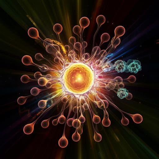
Alkaline pH Promotes NADPH Oxidase-Independent Neutrophil Extracellular Trap Formation: A Matter of Mitochondrial Reactive Oxygen Species Generation and Citrullination and Cleavage of Histone
C. N. D. Souza, L. C. D. Breda, et al.
This study reveals a fascinating connection between pH levels and the formation of NOX-independent neutrophil extracellular traps (NETs). Conducted by Cristiane Naffah de Souza and colleagues, the research uncovers how alkaline pH enhances NET formation through increased calcium influx and mitochondrial reactive oxygen species. The implications for understanding sterile inflammation could lead to new therapeutic approaches.
Related Publications
Explore these studies to deepen your understanding
Adjacent work that informs or extends this paper's methodology and findings.
Medicine and Health
Progression of Cystic Fibrosis Lung Disease from Childhood to Adulthood: Neutrophils, Neutrophil Extracellular Trap (NET) Formation, and NET Degradation
M. A. Khan, Z. S. Ali, et al.

Engineering and Technology
Ultrasonic activation of inert poly(tetrafluoroethylene) enables piezocatalytic generation of reactive oxygen species
Y. Wang, Y. Xu, et al.
Medicine and Health
SK3 channel and mitochondrial ROS mediate NADPH oxidase-independent NETosis induced by calcium influx
D. N. Douda, M. A. Khan, et al.
Medicine and Health
Neutrophil Extracellular Trap Formation: Physiology, Pathology, and Pharmacology
M. Ravindran, M. A. Khan, et al.


