
Medicine and HealthNATURE COMMUNICATIONS
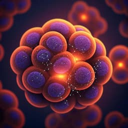
A single-cell nanocoating of probiotics for enhanced amelioration of antibiotic-associated diarrhea
J. Pan, G. Gong, et al.
Dive into groundbreaking research that unveils a novel strategy to protect probiotic bacteria from antibiotics! Conducted by a team that includes Jiezhou Pan and Guidong Gong, this study introduces a cutting-edge 'nanoarmor' that significantly reduces antibiotic-associated diarrhea and promotes better gut health during treatment.
Related Publications
Explore these studies to deepen your understanding
Adjacent work that informs or extends this paper's methodology and findings.
Medicine and Health
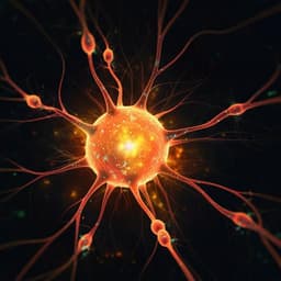
The single-cell transcriptomic atlas iPain identifies senescence of nociceptors as a therapeutical target for chronic pain treatment
P. Techameena, X. Feng, et al.
Medicine and Health
HIDDEN: a machine learning method for detection of disease-relevant populations in case-control single-cell transcriptomics data
A. Goeva, M. Dolan, et al.
Medicine and Health
Single-cell analysis of two severe COVID-19 patients reveals a monocyte-associated and tocilizumab-responding cytokine storm
C. Guo, H. Ma, et al.

Psychology
Factors associated with the outcomes of a novel virtual reality therapy for military veterans with PTSD: Theory development using a mixed methods analysis
B. Hannigan, R. V. Deursen, et al.


