Medicine and Health
Using deep learning to predict abdominal age from liver and pancreas magnetic resonance images
A. L. Goallec, S. Diai, et al.
Aging induces macroscopic and cellular changes across abdominal organs, including the liver and pancreas, increasing vulnerability to conditions such as fibrosis, fatty liver disease, diabetes, and cancer. Biological age, distinct from chronological age, reflects the physiological state of an individual and is a primary driver of age-related diseases. Predicting biological age from organ-specific data can elucidate aging mechanisms and identify risk factors for accelerated aging that may be targets for intervention. While age predictors have been developed for multiple organs and data types, abdominal MRIs (liver and pancreas) had not been leveraged to predict age. The study aims to build and validate the first abdominal age predictor (AbdAge) using deep learning on liver and pancreas MRI images, quantify heritability and genetic architecture of accelerated abdominal aging, and identify non-genetic correlates across biomarkers, clinical phenotypes, diseases, environmental, and socioeconomic factors.
Prior biological age predictors have used diverse modalities: brain and heart MRIs, electrocardiograms, carotid ultrasound, pulse wave analysis, full-body and chest X-rays, eye fundus/OCT imaging, facial features, blood-based biomarkers, DNA methylation, transcriptomics, proteomics, microbiome, and physical activity data. These studies show that biological aging is multidimensional and organ-specific clocks capture different aspects of aging. However, before this work, abdominal MRI imaging of the liver and pancreas had not been employed to predict age, leaving a gap in understanding abdominal organ aging from imaging data.
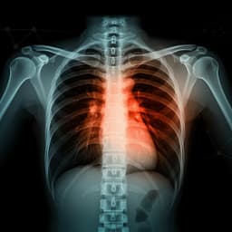
Data: UK Biobank (project ID: 52887) liver MRIs (field 20204) and pancreas MRIs (field 20259) from participants aged 37–82 years. After quality control, 45,552 liver MRIs and 36,784 pancreas MRIs were retained. Demographics included age, sex (genetic where available), and one-hot encoded ethnicity. Image preprocessing: DICOM images (288×384) had low-quality flagged images removed, adaptive histogram equalization applied to generate contrasted versions, legends cropped (resulting in 288×350), and stored as JPG. Both raw and contrasted images were used. Data augmentation: Random vertical/horizontal shifts up to ±10% and rotations up to ±10 degrees applied on-the-fly each epoch. Models: Transfer learning with InceptionV3 and Inception-ResNetV2 (Keras), removing ImageNet top layers as base CNNs. A side fully connected layer (16 nodes) ingested sex and ethnicity and was concatenated with CNN outputs. Post-concatenation dense layers (ReLU, weight decay and dropout regularization; hyperparameters tuned) ended with a linear output for age. Training: Mean squared error loss; RMSE metric; gradient norm clipping (max 1.0); epoch defined as 32,768 images; learning rate halved if training loss plateaued seven epochs; shuffling each pass; model checkpointing on validation improvement; early stopping after 15 epochs without validation loss improvement; jobs capped at 10 hours with resume if improving. Cross-validation: Participants split into 10 folds. Hyperparameters were manually tuned, followed by cross-validation. Ensembling: Three-level hierarchy. Level 1: combine algorithms per dataset (e.g., InceptionV3 and Inception-ResNetV2 on raw liver MRIs). Level 2: combine preprocessing (raw and contrasted) per organ (liver or pancreas). Level 3: combine organ-specific predictors to form AbdAge. Each ensemble was built per fold using elastic nets with inner 10-fold CV on validation folds matched to the testing fold’s model provenance. Performance evaluation: R^2, RMSE, and MAE with uncertainty estimated by (1) standard deviation across folds and (2) bootstrap (1,000 resamples). Bias correction and phenotype definition: Residuals showed age-distribution-induced bias (younger predicted older; older predicted younger). Linear regression of residuals on age was used to correct predictions and residuals. Biological age was the corrected prediction; accelerated aging was the corrected residual (predicted minus chronological age). Genetics: GWAS on average bias-corrected accelerated aging per participant using BOLT-LMM with imputed v3 data; covariates included age, sex, ethnicity, assessment center, and 20 genetic PCs. Autosomes and X chromosome were analyzed in two runs and concatenated (Y excluded). Sample sizes: AbdAge n≈32,475; Liver n≈40,760; Pancreas n≈32,548. Genome-wide significance at p<5e-8. Loci and nearest genes identified using FUMA. Heritability and genetic correlations estimated with BOLT-REML including the X chromosome and the same covariates. Non-genetic correlates (XWAS): Univariate associations of accelerated aging with biomarkers, clinical phenotypes, diseases, family history, environmental, and socioeconomic variables (total 17,459 tests), applying Bonferroni correction (p<0.05 corrected). Additional analyses: Correlations between liver- and pancreas-based accelerated aging and overlap of associated non-genetic variables; correlations and genetic correlations with other organ/tissue aging predictors previously developed by the authors (heart MRI/ECG, musculoskeletal X-ray, carotid ultrasound, brain MRI, eye OCT, physiological measures).
Age prediction performance: Liver MRI model achieved R^2=71.5±0.6%, MAE=3.24±0.04 years, RMSE=4.1±0.05 years. Pancreas MRI model achieved R^2=70.3±0.8%, MAE=3.30±0.04 years, RMSE=4.1±0.04 years. The AbdAge ensemble (combining liver and pancreas) achieved R^2=76.3±0.6%, MAE=2.94±0.03 years, RMSE=3.7±0.03 years. Attention maps: Predictions leveraged not only the target organs (liver or pancreas) but also surrounding abdominal structures, including stomach, spleen, muscle, adipose tissue, bones, and liver (for pancreas images), indicating capture of general abdominal aging features. Heritability and GWAS: Accelerated AbdAge heritability h^2=26.3±1.9%; liver-based h^2=22.3±1.5%; pancreas-based h^2=22.1±1.9%. Genome-wide significant loci: 3 for AbdAge, 2 for Pancreas, and 11 for Liver accelerated age. Notable loci include EFEMP1 (shared between AbdAge and Liver), PLEKHA1/ARMS2/HTRA1 (AbdAge and Pancreas region), PEPD (Pancreas), and multiple loci for Liver (e.g., ADCY3/DNAJC27/EFR3B, TSEN2/C3orf83/MKRN2, BANK1/SLC39A8, WNT16/FAM3C, KLF12, NDRG2/ARHGEF40/ZNF219/TMEM253, RALY). XWAS overview: Of 17,459 tests, 1,456 (8.34%) were significant (average absolute correlation 0.044; range 0.022–0.091; IQR 0.034–0.053). Biomarkers associated with accelerated aging: Strongest categories were body impedance (100% of measures; e.g., right arm 0.056, left arm 0.055, whole body 0.042), blood pressure (66.7%; diastolic 0.050, systolic 0.036), and pulse wave analysis (46.7%; diastolic 0.050, systolic 0.048, mean arterial 0.046). Biomarkers associated with decelerated aging: Hand grip strength (left 0.056; right 0.049), symbol digit substitution performance (0.036; 0.035), and heel bone densitometry (quantitative ultrasound index 0.091; bone mineral density 0.090; speed of sound 0.089). Clinical phenotypes: Accelerated aging linked to general health (overall health rating 0.069; recent weight loss 0.065; long-standing illness/disability 0.050), chest pain (pain walking 0.032; pain ceasing when standing 0.023), and breathing (shortness of breath on level ground 0.031). Decelerated aging linked to sexual factors (later age at first intercourse 0.030) and stable or gained weight (0.032; 0.024). Diseases: Associations with cardiovascular diseases (hypertension 0.058; atrial fibrillation/flutter 0.045; chronic ischaemic heart disease 0.029), general health history (0.046; 0.042; 0.030), and pulmonary diseases (COPD 0.034; asthma 0.026; pleural effusion 0.024). Environmental: Accelerated aging associated with smoking (pack-years proportion 0.090; pack-years 0.086; smoked most/all days 0.066), sun exposure (facial aging self-report 0.039; do not know 0.038; time outdoors in summer 0.036), and alcohol intake (red wine 0.043; champagne/white wine 0.043; beer/cider 0.042). Decelerated aging associated with physical activity (strenuous sports practice 0.078; frequency 0.077; duration 0.076), never smoking (0.073), and diet (cereal intake 0.058; no major dietary changes 0.036; bread 0.030). Socioeconomic: Accelerated aging associated with lack of social/leisure activity (0.033) and renting from local authority/council/housing association (0.028). Decelerated aging associated with higher education (college/university degree 0.048), employment (longer working week 0.044; in paid employment/self-employed 0.043; commuting frequency 0.029), and not receiving attendance/disability/mobility allowance (0.040). Predictability from X modalities: No single X-modal subcategory model explained more than 5% of variance in accelerated abdominal aging. Cross-dimension correlations: Liver- and pancreas-based accelerated aging are phenotypically correlated (0.526±0.005) and genetically correlated (0.863±0.036). Overlap of non-genetic associations between liver and pancreas accelerated aging was high: biomarkers 0.959, clinical 0.926, diseases 0.793, environmental 0.978, socioeconomic 0.969. AbdAge correlated moderately with other biological age dimensions (e.g., heart MRI age 0.45; musculoskeletal 0.35; OCT eye age 0.04; median across 28 phenotypes 0.15). Genetic correlations were highest with musculoskeletal spine aging (0.56), lower with knee (0.23) and hip (0.25).
The study demonstrates that deep convolutional neural networks can accurately predict chronological age from abdominal MRIs, yielding a robust AbdAge measure. Attention maps and the improvement from combining liver and pancreas models indicate that predictions capture general abdominal aging features rather than organ-specific signals. Consequently, liver- and pancreas-based accelerated aging measures are substantially phenotypically and genetically correlated, and show highly overlapping non-genetic associations. Genetic analyses reveal that accelerated abdominal aging is moderately heritable and implicates loci such as EFEMP1 and the PLEKHA1/ARMS2/HTRA1 region, linking AbdAge to pathways shared with other aging-related traits (e.g., age-related macular degeneration) and adiposity/body fat distribution. Non-genetic associations align AbdAge with cardiometabolic and pulmonary health, general health status, and lifestyle factors: smoking and lower physical activity are associated with accelerated aging, whereas greater physical activity and higher education are associated with decelerated aging. Some associations (e.g., inverse relations with BMI/weight) may reflect weight loss in older age or disease states. AbdAge shows only modest correlation with most other organ-specific age predictors, highlighting the multidimensional nature of aging. The findings suggest that while AbdAge reflects global abdominal aging, segmentation-based approaches may be needed for organ-specific liver or pancreas aging assessments. The low variance explained by single X-modalities implies that abdominal aging is influenced by many small-effect factors and complex interactions.
This work introduces the first abdominal age predictor (AbdAge) from liver and pancreas MRIs, achieving high accuracy and revealing that accelerated abdominal aging is moderately heritable and associated with specific genetic loci and broad lifestyle, clinical, and socioeconomic factors. AbdAge provides a quantitative phenotype that may aid in understanding disease risk, stratifying patients, and evaluating interventions. Future research should develop segmentation-based organ-specific predictors, investigate mechanistic links such as stromal stellate cell activity and fibrosis in liver and pancreas, assess causal relationships using Mendelian randomization, and explore AbdAge as an endpoint in clinical trials of rejuvenation therapies. Expanding analyses across diverse ancestries and longitudinal data will further clarify generalizability and temporal dynamics.
The liver- and pancreas-based models capture general abdominal aging rather than organ-specific aging, limiting organ-specific interpretation without segmentation. Predictions required correction for age-distribution-related residual bias. GWAS analyses were restricted to individuals of White ancestry to mitigate population stratification, limiting generalizability. It is unclear whether cellular processes hypothesized to mediate aging (e.g., stellate cell activation) are detectable with the MRIs used. Evidence for shared genetic architecture between AbdAge and other MRI-based aging phenotypes beyond musculoskeletal spine was limited by available data.
Related Publications
Explore these studies to deepen your understanding of the subject.