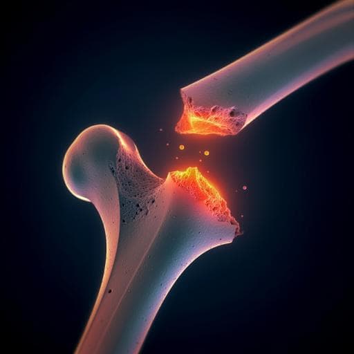
Introduction
Bone's remarkable fracture resistance, or toughness, stems from its hierarchical structure, encompassing nanoscale collagen and mineral phases and microscale osteonal structures. While bone mineral density (BMD) is the primary clinical indicator of fracture risk, it primarily reflects bone's elastic properties and strength. Collagen, however, significantly contributes to post-yield properties and failure energy. Degraded collagen, prevalent in aging and various diseases, increases fracture risk. Fracture resistance arises from intrinsic (material) mechanisms at the nanoscale (e.g., collagen plasticity) and extrinsic mechanisms at the microscale (e.g., crack deflection). This study focuses on the interplay between these mechanisms, particularly how nanoscale collagen damage affects microscale toughening. Synchrotron radiation micro-computed tomography (SRµCT) is an advantageous tool for examining microscale toughening mechanisms, but high-resolution imaging can over-irradiate the bone, altering its properties. To address this, the researchers utilize low-dose imaging combined with deep learning to denoise images and maintain the bone's inherent mechanical properties. The hypothesis is that collagen network alteration decreases crack deflection and reduces bone toughness.
Literature Review
The literature extensively covers the hierarchical structure of bone and its contribution to toughness. Studies have shown that mineral content primarily affects bone strength and elasticity, while collagen is crucial for post-yield behavior and energy absorption. Degraded collagen is linked to increased fracture risk in various conditions, including aging, osteoporosis, and certain diseases. Research highlights both intrinsic (nanoscale, material-based) and extrinsic (microscale, microstructural) toughening mechanisms. Intrinsic mechanisms, primarily influenced by collagen, impede crack initiation and growth, while extrinsic mechanisms, like crack deflection, dissipate energy through microstructural features. Existing research predominantly focuses on the role of microstructural features in extrinsic toughening. The use of SRµCT for studying crack propagation in bone is well established. However, the impact of collagen damage on these microscale mechanisms remains poorly understood. The challenge of over-irradiation during SRµCT imaging is addressed in prior work by employing lower-dose scans and deep learning techniques for image enhancement. This study innovates by combining these techniques for in situ mechanical testing.
Methodology
Bovine bone samples were heat-treated at various temperatures (Room Temperature, 100°C, 130°C, 160°C, 190°C) to induce different levels of collagen damage. A trypsin-hydroxyproline assay quantified the extent of collagen denaturation. Macroscale mechanical properties were assessed using flexural strength tests, measuring ultimate strain, work to fracture, and Young's modulus. In situ SRµCT toughness testing was performed on notched samples. Low-dose SRµCT scans were acquired during three-point bend tests to monitor crack propagation in near real-time. A mixed-scale dense convolutional neural network (CNN) was trained using high- and low-quality reference scans to denoise the low-dose images from the in situ tests. The CNN also performed semantic segmentation to identify the crack path. Digital volume correlation (DVC) was used to track the displacement of osteocyte lacunae and determine the internal strain state during loading. Crack deflection was quantified from the segmented SRµCT images. Statistical analysis (t-tests and ANOVA with Tukey post hoc test) was used to compare different groups.
Key Findings
The trypsin-hydroxyproline assay confirmed significant collagen damage above 130°C. Flexural strength tests revealed that collagen damage primarily affected post-yield properties (ultimate strain and work to fracture), with no significant change in Young's modulus. In situ SRµCT toughness testing showed a substantial reduction (50-70%) in both crack initiation and crack growth toughness in samples with substantial collagen damage (>3%). Crack deflection analysis demonstrated a significant decrease in both the frequency and severity of crack deflections in damaged collagen groups. DVC revealed that the internal strain state influenced crack propagation. In samples with undamaged collagen, strain extended further from the crack tip, leading to more crack deflections. In contrast, damaged collagen samples showed more confined strain around the crack tip, resulting in straighter crack paths.
Discussion
The findings directly address the research question by demonstrating a strong link between nanoscale collagen damage and reduced bone toughness. The reduction in macroscale toughness is directly linked to the impairment of the microscale crack deflection mechanism, a key contributor to bone's ability to resist crack propagation. This highlights a cooperative interaction between intrinsic (collagen-based) and extrinsic (microstructural) toughening mechanisms. The results challenge the traditional focus solely on BMD as an indicator of fracture risk and emphasize the crucial role of collagen quality in bone fragility. These findings are relevant to the field by providing a mechanistic understanding of how collagen damage contributes to bone fragility in diseases and aging. The development of the in situ SRµCT testing technique is a significant advancement, enabling dynamic analysis of crack propagation with damage mitigation.
Conclusion
This study reveals a critical link between nanoscale collagen damage and reduced bone toughness, mediated by the impairment of microscale crack deflection. The novel in situ SRµCT testing technique combining low-dose imaging and deep learning is a powerful tool for investigating bone fracture mechanics. Future studies should investigate the effects of collagen damage from disease processes on bone fracture properties using this technique, potentially providing insights into therapies for bone fragility diseases.
Limitations
The relatively small sample size (particularly in the in situ SRµCT tests) could limit the statistical power of the findings. The heat treatment method, while useful for isolating the effect of collagen damage, might not perfectly mimic the complexities of collagen degradation in disease. Differences between bovine and human bone microstructure (e.g., presence of plexiform bone) could affect the generalizability of findings. While DVC provided valuable insights, there is a potential for some strain exaggeration due to microcracks below the image resolution.
Related Publications
Explore these studies to deepen your understanding of the subject.