
Biology
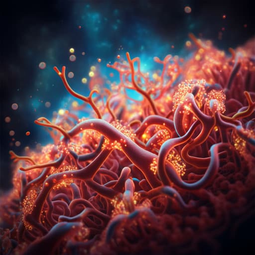
Transplanted human iPSC-derived vascular endothelial cells promote functional recovery by recruitment of regulatory T cells to ischemic white matter in the brain
B. Xu, H. Shimauchi-ohtaki, et al.
This groundbreaking research by Bin Xu, Hiroya Shimauchi-Ohtaki, Yuhei Yoshimoto, Tetsushi Sadakata, and Yasuki Ishizaki reveals how transplanting human iVECs can transform outcomes in ischemic stroke models, particularly by enhancing the recruitment of regulatory T cells and promoting myelin regeneration. Dive into the fascinating interplay between cellular therapies and neuroinflammation!
~3 min • Beginner • English
Introduction
Ischemic stroke, the predominant form of stroke, frequently damages cerebral white matter, leading to demyelination, neuroinflammation, and persistent functional deficits. Following white matter ischemia, activated microglia/macrophages release pro-inflammatory cytokines and proteases that disrupt the blood–brain barrier, facilitating infiltration of peripheral immune cells, including T lymphocytes, which can amplify neuroinflammation and demyelination. Prior work from the authors demonstrated that transplantation of vascular endothelial cells, including those derived from human iPSCs (iVECs), improves locomotor deficits, reduces infarct size, promotes remyelination, and suppresses neuroinflammation, and increases oligodendrocyte precursor cells (OPCs). However, how transplanted endothelial cells modulate neuroinflammation and promote remyelination remained unclear. This study investigates whether the peripheral immune system—particularly regulatory T cells (Tregs)—contributes to the beneficial effects of iVEC transplantation on ischemic white matter injury and functional recovery.
Literature Review
The paper situates the work within evidence that neuroinflammation critically influences ischemic stroke outcomes, with microglial activation, pro-inflammatory cytokines (e.g., TNF-α, IL-1β), and MMPs (e.g., MMP-9) disrupting the BBB and enabling lymphocyte infiltration. Previous studies have shown: (1) endothelial cell transplantation ameliorates ischemic white matter damage and increases OPC survival; (2) endothelial cell-derived extracellular vesicles support OPC survival, proliferation, and motility, partly via fibronectin-mediated uptake; (3) Treg involvement in CNS repair—Tregs suppress neuroinflammation, reduce astrogliosis, and can promote remyelination in multiple models; and (4) pharmacological modulation of lymphocyte trafficking (e.g., fingolimod/FTY720) influences neuroinflammation and microglial polarization. The authors leverage this literature to test whether iVECs act through recruitment of peripheral Tregs to promote remyelination beyond mere suppression of neuroinflammation.
Methodology
- Animals: Male Sprague–Dawley rats, 8 weeks old (250–270 g), housed two per cage, standard conditions. Random assignment to groups; ethical approvals in place.
- White matter infarct model: Endothelin-1 (ET-1) microinjection into the left internal capsule (IC) to induce ischemia. Stereotaxic coordinates: 2.0 mm posterior and 6.6 mm lateral to bregma, 4.6 mm depth; needle angled 30° away from site to minimize mechanical damage and avoid ventricular leakage. ET-1 dose: 1 μl of 100 pmol/μl at 0.2 μl/min; needle left in place 10 min before withdrawal.
- iPSC culture and iVEC differentiation: Human IMR90-4 iPSC line (WiCell) maintained on Matrigel in mTeSR1, passaged with Versene. Differentiation to endothelial cells followed prior protocol: unconditioned medium (UM) for 6 days, then human endothelial serum-free medium (hESFM) with PDS, bFGF (20 ng/ml), and retinoic acid (10 μM) for 48 h; cells replated on collagen IV/fibronectin and maintained in hESFM+PDS. iPSC passages 34–44 were used.
- Human Treg isolation and expansion: PBMCs from healthy donors isolated via density gradient; CD4+CD25+ Tregs purified using MACS. Expanded for 14 days using anti-CD3/CD28 stimulation (MACSiBead particles) in TexMACS medium with human IL-2 (500 U/ml), 5% human AB serum, and β-mercaptoethanol. Post-expansion, flow cytometry confirmed Treg phenotype (CD4+ CD25+ Foxp3+; ~94%).
- Transplantation and treatment timeline: Day 0 ET-1; Day 7 cell transplantation and start FTY720 in applicable groups; assessments at days 10, 14, 21. Transplants delivered stereotaxically at same IC coordinates: 2 μl of iVEC suspension (0.2% BSA/HBSS) at 50,000 cells/μl; or 2 μl of iVEC+Treg mixture (1:1) at 50,000 cells/μl total; or vehicle (0.2% BSA/HBSS). FTY720 administered intraperitoneally at 1 mg/kg daily from days 7–14 to sequester lymphocytes in peripheral lymph nodes.
- MRI: 1 T benchtop scanner (Bruker Icon), T2-weighted imaging at day 7 pre-transplant to confirm lesion size/location; parameters: RARE factor 5, TR 2500 ms, TE 60 ms, in-plane 266×266 μm2, thickness 1250 μm, 5 slices.
- Tissue processing: Perfusion with 4% PFA; cryoprotection in 20% then 30% sucrose; coronal cryosections at 20 μm (120 serial slices/brain) mounted and stored at −80 °C.
- Immunohistochemistry: Blocking with 3% BSA/0.3% Triton X-100; antigen retrieval (CD4/Foxp3: 120 °C, 3 min; Olig2: 120 °C, 20 min, citrate buffer pH 6). Primary antibodies: ED-1 (microglia/macrophages), CD4 (T cells), Foxp3 (Tregs), Olig2 (oligodendrocyte lineage), STEM121 (human cytoplasm). TSA amplification for CD4 and Foxp3. Nuclei stained with Hoechst 33342. Imaging by Zeiss Axioplan2 or Keyence BZ-X710.
- Myelin staining: Luxol fast blue (LFB) to assess remyelination; 12 slices across lesion (200 μm spacing). IC volume computed as percentage of ipsilateral to contralateral IC volume.
- Quantification and statistics: ED-1 fluorescence intensity; counts of CD4+, Foxp3+, and Olig2+ cells. Data as mean ± SEM. Two-tailed Student’s t-test for two-group comparisons; one-way ANOVA with Tukey–Kramer post hoc for multiple groups. Significance at p < 0.05.
- Experimental groups included: Vehicle; iVEC; FTY720; FTY720+iVEC; and, in some experiments, FTY720+iVEC+human Tregs. Replicates generally n = 3–5 per group per time point; experiments repeated three times with similar results.
Key Findings
- iVEC transplantation suppressed neuroinflammation: ED-1+ microglia/macrophages decreased at days 14 and 21 in iVEC-transplanted vs vehicle groups (p < 0.05–0.01), confirming anti-inflammatory effects.
- Reduced CD4+ T-cell infiltration: CD4+ T cells increased over time in vehicle brains but were significantly reduced by iVEC transplantation at days 14 and 21 (p < 0.05), while absent in contralateral intact white matter.
- Increased Tregs with iVECs: Foxp3+ Tregs were scarce at day 7 but increased significantly in iVEC-transplanted lesions at days 10, 14, and 21 vs vehicle (p < 0.05–0.01), indicating recruitment/accumulation of Tregs after iVEC transplantation.
- Peripheral origin of infiltrating T cells: FTY720 (days 7–14) markedly reduced both CD4+ T cells and Foxp3+ Tregs in lesions at day 14 (p < 0.01–0.001), indicating these cells are recruited from the periphery. In iVEC-transplanted animals, FTY720 abrogated the iVEC-induced increase in Tregs (p < 0.001), supporting iVEC-mediated Treg recruitment.
- Neuroinflammation suppression alone did not restore myelin: FTY720 reduced ED-1+ inflammatory cells more strongly than iVECs (p < 0.01–0.001) but did not improve remyelination by LFB analysis, even when combined with iVECs (no significant recovery of damaged IC volume attributable to FTY720 alone).
- iVECs increased oligodendrocyte-lineage cells independent of peripheral lymphocytes: Olig2+ cell numbers increased in iVEC-treated lesions; FTY720 did not alter this increase, indicating the effect on oligodendrocyte lineage amplification is not dependent on peripheral immune cells.
- Tregs promote remyelination without increasing oligodendrocyte-lineage counts: Co-transplantation of human Tregs with iVECs into FTY720-treated animals significantly enhanced remyelination (reduced damaged IC volume; p < 0.001) compared to iVECs alone, while Olig2+ cell numbers were unchanged, suggesting Tregs enhance differentiation/maturation rather than expansion of oligodendrocyte lineage cells.
- Lesion comparability: T2-weighted MRI at day 7 confirmed similar infarct size/location across groups prior to interventions (no significant differences).
Discussion
The study addresses how iPSC-derived vascular endothelial cell transplantation promotes recovery after ischemic white matter injury. Findings show that iVECs suppress neuroinflammation and distinctly modulate adaptive immune infiltration by reducing total CD4+ T cells yet increasing Foxp3+ Tregs in lesions. Using FTY720 to block lymphocyte egress demonstrated that both CD4+ T cells and Tregs in lesions originate from the periphery, and that iVEC-induced Treg accumulation depends on peripheral recruitment. Although neuroinflammation suppression (via FTY720) was robust, it did not translate into remyelination, indicating that dampening inflammation alone is insufficient for myelin repair. Conversely, co-delivering human Tregs with iVECs in a context of reduced endogenous lymphocyte trafficking enhanced remyelination without altering Olig2+ cell numbers, implying that Tregs act to promote oligodendrocyte lineage differentiation and maturation rather than proliferation/expansion. Thus, iVECs likely facilitate myelin repair through two complementary mechanisms: expanding oligodendrocyte-lineage cells (immune-independent) and recruiting Tregs that drive their maturation (immune-dependent). Potential mediators of Treg recruitment by iVECs may include chemokines such as CCL1, CCL20, and CCL22, which the authors propose to investigate. The results refine understanding of cell-based therapies for white matter stroke by highlighting the specific, beneficial role of Tregs in remyelination beyond general immunosuppression.
Conclusion
Transplantation of human iPSC-derived vascular endothelial cells into ischemic white matter reduces neuroinflammation, recruits peripheral Foxp3+ regulatory T cells, and promotes remyelination. Neuroinflammation suppression alone (e.g., by FTY720) does not restore myelin, whereas Treg presence is critical for remyelination, likely by enhancing differentiation/maturation of oligodendrocyte lineage cells rather than increasing their numbers. These findings suggest therapeutic strategies combining endothelial cell transplantation with approaches to recruit or deliver Tregs to optimize white matter repair after stroke. Future studies should identify the iVEC-derived factors mediating Treg recruitment and delineate Treg-driven mechanisms of oligodendrocyte maturation, as well as evaluate broader immune responses (e.g., B cells) and long-term functional outcomes.
Limitations
- Mechanistic details remain unresolved: the specific factors by which iVECs recruit Tregs are not identified, and how Tregs promote remyelination (e.g., differentiation/maturation pathways) is undetermined.
- Immune scope: The study did not examine B-cell responses after iVEC transplantation, which are reported to be influenced by CD4+ T cells after stroke.
- Model and timeframe: Findings are based on a rat ET-1-induced white matter infarct model with assessments primarily between days 10–21; longer-term outcomes and generalizability to other models/species or to human pathology require further investigation.
Related Publications
Explore these studies to deepen your understanding of the subject.