Medicine and Health
The one-step fabrication of porous hASC-laden GelMa constructs using a handheld printing system
S. Jo, J. Lee, et al.
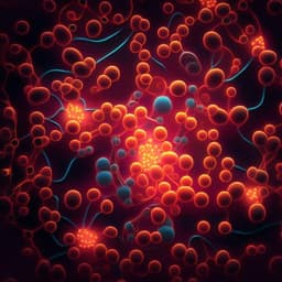
Bioprinted cell-laden hydrogels often suffer from low porosity and limited nutrient and oxygen transport, causing central necrosis in thick, non-porous struts. Prior reports indicate that hydrogel struts thicker than ~200 µm can lead to central cell death and that introducing porosity increases viability. Efficient vascular infiltration is also hindered by non-porous matrices. Consequently, there is a need for a simple, safe, and controllable method to fabricate highly porous, cell-laden constructs that support cell viability and tissue integration. This study addresses that need by introducing a one-step, handheld 3D printing system that simultaneously mixes clean air with a GelMa bioink containing hASCs via inline mesh filters to generate air bubbles, enabling high-porosity constructs without sacrificial porogens or complex equipment. The research evaluates how processing parameters govern pore size and foamability and tests the in vitro activity of hASCs and in vivo functional regeneration in a mouse volumetric muscle loss model.
Multiple strategies have been explored to generate porous cell-laden constructs: (1) Sacrificial porogens such as gelatin beads dispersed in alginate, later leached at 37 °C, yield ~70% porosity but require homogeneous bead distribution and multi-step processing. (2) Protein-based whipping approaches (e.g., collagen, GelMa with PVA/LAP) create macro/microporous foams supporting cell viability and vascularization, but require surfactants, vigorous mixing, and careful post-mixing with cells, limiting in situ feasibility. (3) Air injection via syringe can produce GelMa foams with hASCs but is typically a two-step process with challenging pore size control. (4) Aqueous two-phase emulsions (ATPE) enable homogeneous viable cell distribution but have limited control over pore size range and distribution. (5) Gas foaming (e.g., H2 with gelatin/alginate) offers adjustable micropores but involves sacrificial components and extensive washing. (6) Microfluidic N2 foaming yields interconnected, injectable porous hydrogels but requires multi-inlet control and lengthy crosslinking. Table 1 summarizes these methods with their pore sizes, cell types, crosslinking conditions, advantages, and disadvantages, highlighting the need for a simple, sacrificial-free, and in situ-capable approach with tunable porosity.
Bioink synthesis and rheology: GelMa was synthesized by methacrylation of gelatin (porcine skin; 300 g Bloom) in PBS with methacrylic anhydride at 50 °C, dialyzed (MWCO 1000 kDa) at 40 °C for 7 days, and lyophilized. Lyophilized GelMa was dissolved in PBS with 0.3 mg/mL LAP to prepare 5, 10, and 15% w/v solutions. Rheology was characterized using a cone-and-plate rheometer (1 Hz, 1% strain) with temperature sweeps (10–40 °C) and time sweeps under UV doses (0.5–3 J/cm²) to assess photo-crosslinking. The degree of methacrylation used in experiments was ~87.6%. Handheld printing system: A homemade handheld dual-syringe device (commercial clamp gun base) delivered GelMa+cells and air into a three-way stopcock and through inline polyethylene mesh filters to create bubbles, ejecting porous bioink via a 16 G tapered nozzle. Filter sizes tested: FS-1 (274.5 ± 26.0 µm), FS-2 (196.3 ± 26.4 µm), FS-3 (121.5 ± 43.9 µm). Number of filters (FN) varied (0–3). Processing parameters: bioink temperature (15 vs 33 °C), GelMa concentration (5, 10, 15% w/v), UV dose (0–3 J/cm²), filter size/number, bioink:air mixing ratios (1:1, 1:2, 1:3, 1:4), and flow rate (3, 6, 12 mL/s) were systematically evaluated. Unless stated otherwise, optimized conditions used were: 10% w/v GelMa at 33 °C, FS-3 with FN=3, bioink:air 1:3, flow rate 3 mL/s, and UV dose 1 J/cm². Characterization: Optical microscopy and SEM assessed pore morphology. Pore sizes (n=4 samples, 600 measurements across top/bottom) and pore size coefficient of variation were quantified by ImageJ. Foamability (%) = Vfoam/Vtotal×100 was measured (n=8). In vitro assays: Porous and non-porous constructs with hASCs (1×10^7 cells/mL) or C2C12 were fabricated in PDMS molds (8 mm diameter, 10 mm height). Viability was assessed by calcein AM/ethidium homodimer live/dead staining and confocal imaging; proliferation by MTT at days 1, 3, 7; cytoskeleton by DAPI/phalloidin with F-actin area quantification; myogenic differentiation by MHC immunofluorescence (MF20 primary, Alexa Fluor 488 secondary) and positive index quantification. In vivo mouse VML model: Ten-week-old male C57BL/6 mice were assigned to five groups (n=3/group): sham, defect (no treatment), acellular foam (porous GelMa without cells), hASC-printed (conventional bioprinted hASC-laden GelMa), and hASC-foam (porous hASC-laden GelMa via handheld in situ). Approximately 40% of the TA muscle was excised; constructs were implanted and UV-crosslinked in situ (1 J/cm²). Group-specific formulations: Porous GelMa [foam]: GelMa 5% w/v + hASCs (1×10^7 cells/mL) + LAP 0.3 w/v%; Normal GelMa [printed]: GelMa 10% w/v + hASCs (1×10^7 cells/mL) + LAP 0.3 w/v%. Functional outcomes: hindlimb grip strength (weeks 1–4), latency to fall (rotarod, max 300 s), and TA muscle weight at week 4. Histology and immunofluorescence at 4 weeks: H&E and Masson's Trichrome to assess newly formed muscle fiber area, myofiber diameter, and fibrotic area; IF for MHC, MRPL11, HLA-A, and LAMA1 with quantitative image analysis.
- Porous architecture: Handheld in situ air-bubbling and filtering produced hASC-laden GelMa constructs with macropores and interconnected micropores. Typical pore size ~350 ± 240 µm; porosity up to ~97%; foamability up to ~93%.
- Cell viability and safety: Live/dead assays showed high viability in porous constructs (≈98.3% in GelMa foam). UV crosslinking reduced viability above 1 J/cm²; viability ~90% at ≤1 J/cm², decreasing with higher doses (2–3 J/cm²).
- Rheology/temperature: At 33 °C (sol-like, lower viscosity) large bubbles broke into smaller ones across filters; at 15 °C (gel-like, higher viscosity) bubbles remained larger and less stable.
- GelMa concentration: At 33 °C with FS-3 and bioink:air 1:3, pore sizes (mean) decreased with lower viscosity: 5% 393.9 µm; 10% 349.8 µm; 15% 295.5 µm, but 15% had the widest size distribution (higher coefficient of variation ~0.84 vs 0.45–0.54). Foamability (%) was lowest for 5% (86.36%) and higher for 10% (96.85%) and 15% (97.66%). C2C12 viability: 97.32% (5%), 98.58% (10%), 85.18% (15%). 10% GelMa at 33 °C selected.
- Filter size/number: Without filters, pores were very large (1413.2 ± 1190 µm) and foamability low. Smaller mesh (FS-3, 121.5 ± 43.9 µm) and increased filter number (up to 3) reduced pore size (to ~360–500 µm) and increased foamability; effects saturated beyond three filters, and smaller meshes required excessive pressure.
- Bioink:air ratio: Increasing air fraction from 1:1 to 1:3 raised foamability from ~67% to ~93%, with little further gain at 1:4; pore size differences among 1:2–1:4 were not significant.
- Flow rate: With FS-3 and 1:3 ratio, pore sizes at 3, 6, 12 mL/s were 349.8, 341.8, and 319.3 µm; foamability ~95.60%, 94.26%, 94.10%. Cell viability decreased with higher flow (likely increased shear): ~98.58% (3 mL/s), ~86.22% (6 mL/s), ~82.56% (12 mL/s). Flow set to 3 mL/s.
- In vitro performance: Porous constructs (air volume fraction ~65%; density 0.25 g/mL; porosity 97%) showed significantly higher hASC proliferation (MTT) than non-porous controls. Regional viability at day 7: bottom region 54.11% (control) vs 88.75% (porous), indicating improved nutrient/metabolite transport. F-actin staining showed more spread cytoskeleton in porous constructs; MHC-positive index was significantly higher in porous vs control, indicating enhanced myogenic differentiation.
- In situ feasibility: Handheld device enabled rapid deposition and UV crosslinking (~2 s coverage) directly on VML defects.
- In vivo regeneration (4-week mouse VML): hASC-foam group exhibited significantly greater grip strength and latency to fall than defect, acellular foam, and hASC-printed groups; muscle weight in hASC-foam most closely matched sham. Histology showed newly formed muscle fiber area and myofiber diameter in hASC-foam comparable to sham, with significantly reduced fibrotic area vs defect and hASC-printed. IF markers (MHC, MRPL11, HLA-A, LAMA1) were strongly positive in hASC-foam, higher than other treated groups and comparable (for MHC area) to sham, indicating survival and myogenic differentiation of hASCs within porous constructs.
This study directly addresses the major limitation of conventional cell-laden hydrogel constructs—central hypoxia and necrosis due to low porosity—by introducing a one-step, in situ, handheld printing strategy that generates air-bubbled, highly porous GelMa matrices laden with hASCs. By tuning bioink rheology, filter size/number, bioink–air ratio, UV dose, and flow rate, the system reproducibly achieved high foamability and macroporous structures while maintaining high cell viability. The porous constructs enhanced nutrient and oxygen transport, as evidenced by substantially higher cell viability in the bottom regions, greater proliferation, more developed F-actin cytoskeleton, and increased myogenic differentiation (MHC positivity) compared to non-porous controls. Importantly, in vivo application in a VML model demonstrated superior functional recovery and histological regeneration with hASC-laden porous constructs over acellular foam and conventionally bioprinted (non-porous) constructs. These findings underscore the significance of in situ-generated macroporosity for vascular and tissue integration and validate the handheld approach as a practical, rapid, and equipment-light method suitable for translational scenarios where onsite fabrication is advantageous.
A lightweight handheld 3D printing system that co-injects air and hASC-laden GelMa through inline mesh filters enables one-step fabrication of highly porous constructs (porosity ~97%, pore size ~350 µm, foamability ~93%) with high cell viability and enhanced myogenic activity. Optimized parameters (10% GelMa at 33 °C, FS-3 filters ×3, bioink:air 1:3, 3 mL/s, UV 1 J/cm²) yielded constructs that improved in vitro proliferation and differentiation and significantly accelerated functional and histological regeneration in a mouse VML model compared to non-porous controls. This approach removes the need for sacrificial porogens and complex bioprinters, facilitating in situ application. Future work should focus on precise control of pore size distributions, long-term and large-animal studies, standardization of in situ workflows, integration with angiogenic/neurogenic cues, and extension to other tissues and bioinks.
Pore size and porosity could not be precisely controlled; effects saturated beyond three filters and smaller meshes demanded high pressure. Elevated UV doses compromised cell viability, and higher viscosity (15% GelMa) or flow rates reduced viability due to shear stress. In vivo comparisons used different GelMa concentrations between groups (foam: 5% vs printed: 10%), potentially confounding mechanical and biological outcomes. The in vivo study was limited to a small sample size (n=3 per group) and a 4-week endpoint in a mouse model, which may limit generalizability. Handheld operation may introduce user-dependent variability.
Related Publications
Explore these studies to deepen your understanding of the subject.