Medicine and HealthNature Communications
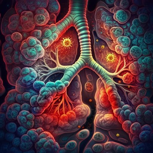
The establishment of COPD organoids to study host-pathogen interaction reveals enhanced viral fitness of SARS-CoV-2 in bronchi
L. L. Y. Chan, D. E. Anderson, et al.
This groundbreaking study by Louisa L. Y. Chan and colleagues uncovers crucial insights into chronic obstructive pulmonary disease (COPD) through novel organoid models. By establishing nasopharyngeal and bronchial organoids from healthy and COPD patients, researchers reveal significant alterations in cellular behavior and heightened inflammatory responses to infections, providing a new perspective on host-pathogen interactions in the lungs.
Related Publications
Explore these studies to deepen your understanding
Adjacent work that informs or extends this paper's methodology and findings.

Medicine and Health
Risk factors for and pregnancy outcomes after SARS-CoV-2 in pregnancy according to disease severity: A nationwide cohort study with validation of the SARS-CoV-2 diagnosis of Nordic Federation of Societies of Obstetrics and Gynecology (NFOG)
A. J. M. Aabakke, T. G. Petersen, et al.
Medicine and Health
Broad host range of SARS-CoV-2 and the molecular basis for SARS-CoV-2 binding to cat ACE2
L. Wu, Q. Chen, et al.

Medicine and Health
Evolution of the SARS-CoV-2 spike protein in the human host
A. G. Wrobel, D. J. Benton, et al.

Medicine and Health
Molecular interaction and inhibition of SARS-CoV-2 binding to the ACE2 receptor
J. Yang, S. J. L. Petitjean, et al.


