
BiologyNature Communications
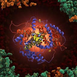
Structure of the native pyruvate dehydrogenase complex reveals the mechanism of substrate insertion
J. Škerlová, J. Berndtsson, et al.
This groundbreaking research reveals the intricate workings of the pyruvate dehydrogenase complex in *E. coli*, showcasing a cryo-EM reconstruction that uncovers the assembly and dynamics of dihydrolipoyl transacetylase. Conducted by Jana Škerlová, Jens Berndtsson, Hendrik Nolte, Martin Ott, and Pål Stenmark, this study sheds light on the active site's substrate shuttling mechanism.
Related Publications
Explore these studies to deepen your understanding
Adjacent work that informs or extends this paper's methodology and findings.
Medicine and Health
The complex structure of GRL0617 and SARS-CoV-2 PLpro reveals a hot spot for antiviral drug discovery
Z. Fu, B. Huang, et al.

Medicine and Health
The cryo-EM structure of the bd oxidase from *M. tuberculosis* reveals a unique structural framework and enables rational drug design to combat TB
S. Safarian, H. K. Opel-reading, et al.
Biology
Key role of quinone in the mechanism of respiratory complex I
J. Gutiérrez-fernández, K. Kaszuba, et al.

Medicine and Health
Crystal structure of adenosine A<sub>2A</sub> receptor in complex with clinical candidate Etrumadenant reveals unprecedented antagonist interaction
T. Claff, J. G. Schlegel, et al.


