
Medicine and Health
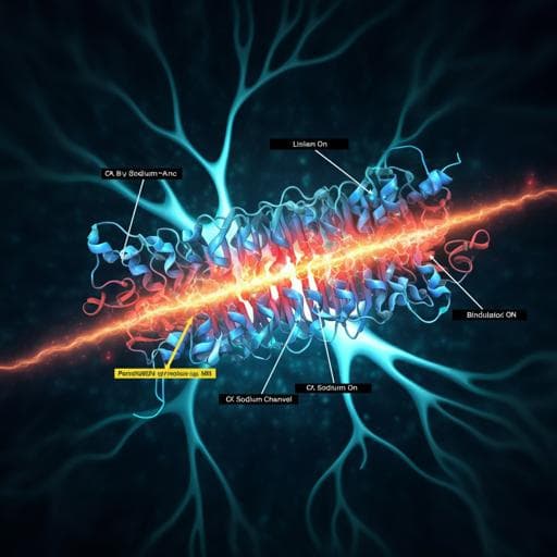
Structural basis for modulation of human Nav1.3 by clinical drug and selective antagonist
X. Li, F. Xu, et al.
Dive into the fascinating world of voltage-gated sodium channels! This study reveals how NaV1.3 interacts with specific modulators like bulleyaconitine A and ICA121431, shedding light on their unique binding mechanisms. Conducted by esteemed researchers including Xiaojing Li and Feng Xu, these findings are paving the way for targeted therapeutic advancements.
~3 min • Beginner • English
Introduction
Voltage-gated sodium channels (NaV) initiate and propagate action potentials and have nine mammalian isoforms with tissue-specific expression. NaV1.3 (encoded by SCN3A) is highly expressed in the CNS and is implicated in neuronal development, hormone secretion, pain perception, and neurological disorders including epilepsy and polymicrogyria. NaV1.3 is re-expressed in injured sensory neurons, contributing to neuropathic pain, making it a therapeutic target. While many anesthetic/anti-arrhythmic drugs bind the central pore of NaVs, the conserved pore hinders isoform-selective drug development. Distinct modulatory sites exist for natural toxins and small molecules. Site-2 neurotoxins (e.g., aconitine analog bulleyaconitine A, BLA) modulate activation/inactivation and reduce peak current amplitude, but detailed binding and mechanisms remain unclear. Aryl sulfonamides (e.g., ICA121431, ICA) selectively inhibit NaV1.3/NaV1.1 with nanomolar potency by binding within voltage sensor domains, yet the structural basis for ICA selectivity and inhibition of NaV1.3 was unresolved. This study aims to determine high-resolution structures of human NaV1.3/β1/β2 bound to BLA and ICA and to elucidate their distinct modulatory mechanisms.
Literature Review
Prior structural work established conserved architecture of mammalian NaVs, with α-subunits comprising four domains (I–IV) each with a voltage-sensor (S1–S4) and pore module (S5–S6). Auxiliary β1–β4 subunits modulate kinetics and expression. Multiple toxin and drug binding sites have been mapped, including pore blockers (local anesthetics/anti-arrhythmics) near S6 and gating-modifier toxins acting on VSDs. Site-2 neurotoxins (aconitine, veratridine, grayanotoxin, batrachotoxin) shift activation to more negative voltages and alter inactivation/selectivity; mutagenesis implicated DI/DII S6 residues in their binding but with diverse modes. Aryl sulfonamide antagonists (e.g., ICA121431, GX-936, PF-04856264) bind within VSDs and show isoform selectivity, with prior chimeric structures and mutagenesis indicating interactions with S4 gating charges. However, the precise structural determinants for BLA and ICA binding in human NaV1.3, and the allosteric mechanisms underlying their modulation, required direct visualization.
Methodology
- Heterologous expression and electrophysiology: Human NaV1.3 with β1 and β2 were co-expressed in HEK293 cells. Whole-cell voltage-clamp recordings assessed activation and steady-state fast inactivation (20-ms depolarizations from -80 to 10 mV; pre-pulses for inactivation). Use-dependent protocols (1000 pulses at 5 Hz) probed BLA effects; inactivated-state protocols (pre-pulse at -50 mV for 8 s) probed ICA inhibition. Dose-response for ICA was determined under inactivated-state conditions. Drugs were applied via puffing in bath solution.
- Protein expression and purification: Human NaV1.3 splice variant 2 (Q9NY46-2), β1 (Q07699), β2 (O60939) were cloned into BacMam vectors with C-terminal mCherry and Twin-Strep on NaV1.3. Baculoviruses produced in Sf9 infected HEK293F for expression. Membranes were solubilized (DDM/CHS), purified via Strep-Tactin affinity and size exclusion chromatography in GDN. Ligands were included throughout purification (ICA: up to 500 µM; BLA: 50–200 µM).
- Cryo-EM: Samples vitrified on Quantifoil grids, data collected on Titan Krios (300 kV) with Gatan K2 Summit and GIF. Super-resolution movies at 1.04 Å/pixel, defocus −1.2 to −2.2 µm, total dose ~60 e/Ų. Motion correction (MotionCor2), CTF estimation (Gctf), particle processing (RELION 3.0), final refinement (cisTEM or cryoSPARC). Final reconstructions at 3.3 Å (NaV1.3/β1/β2-BLA) and 3.4 Å (NaV1.3/β1/β2-ICA).
- Model building: Homology placement from human NaV1.2 α and NaV1.7 β subunits (PDBs 6J8E, 6J8H), manual refinement in Coot, Phenix refinement. Ligand densities interpreted for BLA (central cavity near DI–DII fenestration) and ICA (VSDIV extracellular cleft), and interacting residues identified. Pore radius calculated (HOLE). Comparative structural analyses with NaVAb open/resting and NaV1.5 LqhIII-bound structures.
Key Findings
- Structures: Determined cryo-EM structures of human NaV1.3/β1/β2 bound to bulleyaconitine A (BLA) at 3.3 Å and ICA121431 (ICA) at 3.4 Å. All VSDs are in activated conformations and the activation gate is non-conductive; the IFM inactivation motif is engaged, indicating inactivated state.
- Electrophysiology baseline: NaV1.3 exhibits V1/2 of activation −18.8 ± 0.4 mV (n=17) and steady-state fast inactivation V1/2 −45.4 ± 1.2 mV (n=13).
- BLA binding and function: BLA produces use-dependent inhibition; at 50 µM, ~90% current blocked after 1000 pulses at 5 Hz, with minimal effect on the first pulse. BLA induces a small persistent current (<5% of peak) at −50 mV. Structural density places BLA in the central cavity near the DI–DII fenestration, tightly coordinated by P-loops and S6 of DI/DII with five H-bonds to T381, C941, G942 (carbonyls) and Q382, N972 (sidechains). Receptor site residues include M380, T381, V416, I419, L420 (DI) and C941, G942, M968, N972, L976 (DII). BLA is 8.1 Å and 4.3 Å from NaV1.5 pore-blocker residues F1759 and Y1766, respectively, indicating a distinct site from local anesthetics. Mechanistically, BLA likely accesses via the open activation gate, stabilizes DI/DII S6 in an open-like conformation, interacts with P-loops beneath the DEKA selectivity filter, and partially occludes the ion path, explaining reduced peak current with increased open probability and altered selectivity.
- ICA binding, potency, and selectivity: ICA shows strong inactivated-state inhibition with IC50 95.5 ± 9.3 nM and negligible block in resting-state protocols. Density reveals ICA in the extracellular cleft of VSDIV, with the anionic thiazole headgroup engaging S4 gating charges R2–R4 via electrostatics, and additional contacts: G1603S3, A1626S4 (space for headgroup), L1563S2 and M1604S3 (benzene ring), R1560S2 and F1605S3 (diphenyl tail). A proximal lipid density may stabilize the hydrophobic tail. Comparison to GX-936 in NaV1.7 indicates conserved headgroup–gating charge interactions determine potency, while isoform selectivity arises from differences at positions S1559 and R1560 in NaV1.3 (clashing Y1537/W1538 in NaV1.7). Mutations S1559Y and R1560W reduce ICA potency 9-fold and 33-fold, respectively.
- State preference and allosteric mechanism: ICA preferentially binds VSDIV in its activated conformation; binding to deactivated or resting VSD is unfavorable due to loss of R2/R3 interactions and potential clashes with R1. Stabilization of activated VSDIV by ICA promotes IFM motif engagement and maintains the activation gate closed, providing an allosteric inhibition mechanism. In contrast, α-scorpion toxins trap VSDIV in a deactivated state to oppose inactivation and enhance opening.
- Disease mapping: Several disease-associated NaV1.3 mutations map to S4–S5 linkers and S6 regions implicated in gating (e.g., L247P loss-of-function; I875T, P1333L gain-of-function; V1769A persistent current), providing structural context.
Discussion
The structures elucidate how two chemically distinct modulators affect NaV1.3 through separate receptor sites. BLA binds within the conserved central cavity near the DI–DII fenestration, stabilizing DI/DII S6 in an open-like arrangement while physically narrowing the permeation pathway and engaging P-loops below the DEKA locus. This dual action explains reduced peak current alongside increased open probability and altered selectivity characteristic of site-2 neurotoxins. In contrast, ICA binds within the extracellular cleft of VSDIV, with its anionic headgroup forming strong electrostatic interactions with S4 gating charges R2–R4. By preferentially stabilizing activated VSDIV, ICA promotes IFM motif docking and inactivation, leading to pore closure and potent inhibition. The headgroup–gating charge interactions underpin potency across aryl sulfonamides, while residues S1559 and R1560 in NaV1.3 shape a selective pocket for ICA’s hydrophobic core and tail, accounting for NaV1.3/NaV1.1 selectivity relative to NaV1.7. These findings highlight allosteric inhibition via VSDIV as a route to isoform selectivity, in contrast to the highly conserved pore where selective blockade is difficult. The structural mapping of disease mutations further links gating elements to pathophysiology. Overall, the work advances understanding of NaV activation/inactivation coupling, toxin modulation, and avenues for selective NaV therapeutics.
Conclusion
This study reports high-resolution cryo-EM structures of human NaV1.3/β1/β2 bound to bulleyaconitine A and ICA121431, defining distinct binding sites and mechanisms. BLA occupies a receptor site in the central cavity near the DI–DII fenestration, stabilizing open-like S6 conformations and partially occluding the pore, thereby reducing peak current while increasing open probability. ICA binds within the extracellular cleft of activated VSDIV, engaging S4 gating charges to stabilize inactivation and allosterically close the activation gate, with S1559 and R1560 conferring isoform selectivity. These insights provide structural frameworks for designing NaV modulators, particularly isoform-selective VSD-targeted inhibitors. Future work should explore ligand access pathways and dynamics across channel states, lipid contributions to VSD drug binding, extend analyses to other isoforms and aryl sulfonamides, and assess in vivo efficacy and safety to guide therapeutic development.
Limitations
- Structural snapshots capture NaV1.3 in an inactivated state with activated VSDs; dynamic transitions (e.g., ligand access via open gate vs fenestrations) are inferred but not directly observed.
- The β2 subunit shows weak and fragmented density, suggesting mobility or weak binding; its functional contributions may be underrepresented structurally.
- BLA binding was characterized in NaV1.3; while key residues are conserved, differences among isoforms and among diverse site-2 toxins may yield varied binding modes not fully resolved here.
- ICA selectivity determinants are identified for NaV1.3/NaV1.1 versus NaV1.7 by structural comparison and prior mutagenesis; comprehensive mutational validation across all isoforms and in cellular/physiological contexts is beyond the scope.
- Experiments were performed in heterologous systems with a specific NaV1.3 splice variant, which may differ from native cellular environments.
Related Publications
Explore these studies to deepen your understanding of the subject.