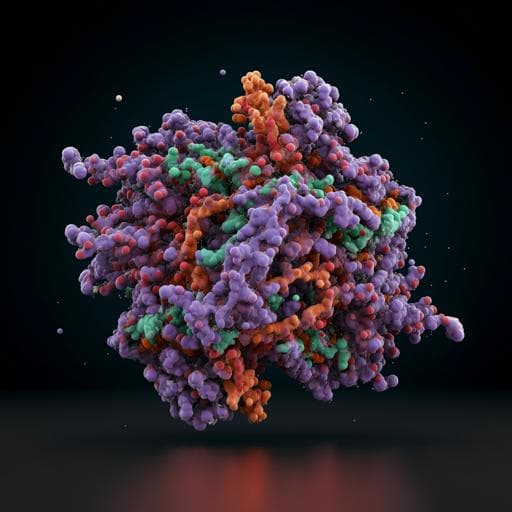
Structural and biochemical mechanism for increased infectivity and immune evasion of Omicron BA.2 variant compared to BA.1 and their possible mouse origins
Y. Xu, C. Wu, et al.
Explore these studies to deepen your understanding
Adjacent work that informs or extends this paper's methodology and findings.

Evaluating the effectiveness of the Kidogo model in empowering women and strengthening their capacities to engage in paid labor opportunities through the provision of quality childcare: a study protocol for an exploratory study in Nakuru County, Kenya
K. Okelo, M. Nampijja, et al.

SARS-CoV-2 disease severity and transmission efficiency is increased for airborne compared to fomite exposure in Syrian hamsters
J. R. Port, C. K. Yinda, et al.

Is it possible for people to develop a sense of empathy toward humanoid robots and establish meaningful relationships with them?
E. Morgante, C. Susinna, et al.

Risk factors for and pregnancy outcomes after SARS-CoV-2 in pregnancy according to disease severity: A nationwide cohort study with validation of the SARS-CoV-2 diagnosis of Nordic Federation of Societies of Obstetrics and Gynecology (NFOG)
A. J. M. Aabakke, T. G. Petersen, et al.


