
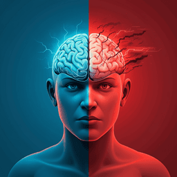
Sleep-like cortical dynamics during wakefulness and their network effects following brain injury
M. Massimini, M. Corbetta, et al.
Explore these studies to deepen your understanding
Adjacent work that informs or extends this paper's methodology and findings.
Prolonged exertion of self-control causes increased sleep-like frontal brain activity and changes in aggressivity and punishment
E. Ordali, P. Marcos-prieto, et al.
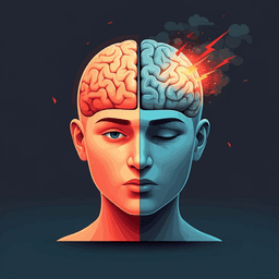
Prolonged exertion of self-control causes increased sleep-like frontal brain activity and changes in aggressivity and punishment
E. Ordali, P. Marcos-prieto, et al.

Prevalence and network structure of post-traumatic stress symptoms and their association with suicidality among Chinese mental health professionals immediately following the end of China's Dynamic Zero-COVID Policy: a national survey
P. Chen, L. Zhang, et al.
Brain mitochondrial diversity and network organization predict anxiety-like behavior in male mice
A. M. Rosenberg, M. Saggar, et al.


