
Medicine and HealthNature Communications
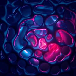
Self-reporting photodynamic nanobody conjugate for precise and sustainable large-volume tumor treatment
Y. Chen, T. Xiong, et al.
Discover the innovative development of MNB-Pyra Nbs, a groundbreaking photodynamic conjugate that effectively suppresses tumors. This pioneering research by Yingchao Chen, Tao Xiong, Qiang Peng, Jianjun Du, Wen Sun, Jiangli Fan, and Xiaojun Peng showcases how these photosensitizers can be activated for self-reporting PDT, making a significant impact in cancer therapy.
Related Publications
Explore these studies to deepen your understanding
Adjacent work that informs or extends this paper's methodology and findings.
Engineering and Technology
Mechanochromic and thermally reprocessable thermosets for autonomic damage reporting and self-healing coatings
S. Yoon, J. H. Choi, et al.
Medicine and Health
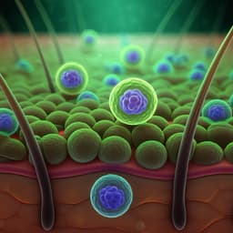
Advances in Photodynamic Therapy for the Treatment of Actinic Keratosis and Nonmelanoma Skin Cancer: A Narrative Review
A. S. Farberg, W. Justin, et al.
Medicine and Health
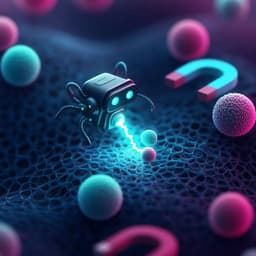
Water-powered self-propelled magnetic nanobot for rapid and highly efficient capture of circulating tumor cells
R. D. Wavhale, K. D. Dhobale, et al.

Medicine and Health
Mindfulness and self-regulation intervention for improved self-neglect and self-regulation in diabetic older adults
M. Motamed-jahromi, M. H. Kaveh, et al.


