
ChemistryCommunications Chemistry
Sample illumination device facilitates in situ light-coupled NMR spectroscopy without fibre optics
J. E. Bramham and A. P. Golovanov
Discover how NMRtorch, a groundbreaking device designed by Jack E. Bramham and Alexander P. Golovanov, illuminates liquid-state NMR samples more effectively than ever. This innovative tool enhances photo-NMR applications, offering insights into photoisomerisation and UV degradation studies.
Related Publications
Explore these studies to deepen your understanding
Adjacent work that informs or extends this paper's methodology and findings.

Physics
Electronic transport driven by collective light-matter coupled states in a quantum device
F. Pisani, D. Gacemi, et al.

Physics
In situ electron paramagnetic resonance spectroscopy using single nanodiamond sensors
Z. Qin, Z. Wang, et al.
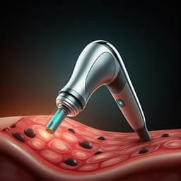
Medicine and Health
Miniaturized all-in-one microneedle device for point of care light therapy
H. Zhao, X. Wang, et al.

Engineering and Technology
A Tuneable Telecom Wavelength Entangled Light Emitting Diode Deployed in an Installed Fibre Network
Z. Xiang, J. Huwer, et al.


