Medicine and HealthNature Communications
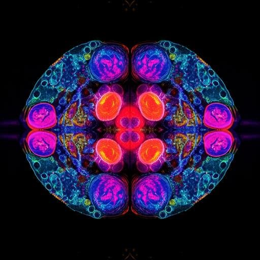
Quantum Microscopy of Cells at the Heisenberg Limit
Z. He, Y. Zhang, et al.
Discover the groundbreaking advancements in quantum microscopy by coincidence (QMC) achieved by Zhe He and colleagues. This innovative technique utilizes entangled photons to break the barriers of super-resolution imaging, allowing for non-destructive bioimaging with a resolution of 1.4 µm in cancer cells. Experience imaging at the Heisenberg limit with unparalleled speed and contrast-to-noise ratio!
Related Publications
Explore these studies to deepen your understanding
Adjacent work that informs or extends this paper's methodology and findings.

Physics
Spooky action at a global distance: analysis of space-based entanglement distribution for the quantum internet
S. Khatri, A. J. Brady, et al.

Physics
Beyond the standard quantum limit for parametric amplification of broadband signals
M. Renger, S. Pogorzalek, et al.

Education
The concept of inclusive education from the point of view of academics specialising in special education at Saudi universities
A. Madhesh

Humanities
Reframing the narrative of magic wind in Arthur Waley's translation of *Journey to the West*: another look at the abridged translation
F. (. Wang, K. Liu, et al.


