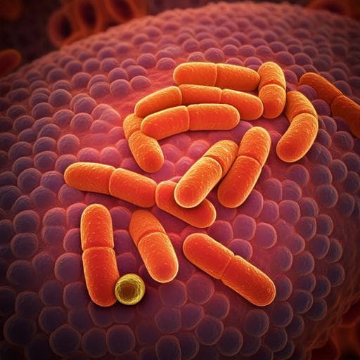
Protection against DSS-induced colitis in mice through FcεRla deficiency: the role of altered *Lactobacillus*
Y. Yin, R. Wang, et al.
Explore these studies to deepen your understanding
Adjacent work that informs or extends this paper's methodology and findings.

Social Networks Play a Complex Role in HIV Prevention Knowledge, Attitudes, Practices, and the Uptake of PrEP Through Transgender Women Communities Centered Around Three "Casas Trans" in Lima, Peru: A Qualitative Study
T. Temelkovska, K. Moriarty, et al.

Exploring the green edge: the role of market orientation and knowledge management in achieving competitive advantage through creativity
Z. Zhang

The mode of action of plant-associated Burkholderia against grey mould disease in grapevine revealed through traits and genomic analyses
Q. Esmaeel, C. Jacquard, et al.
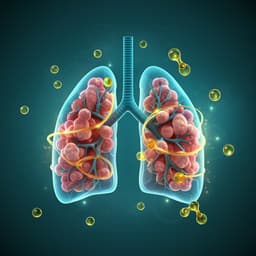
Omega-3 polyunsaturated fatty acids ameliorate PM2.5 exposure induced lung injury in mice through remodeling the gut microbiota and modulating the lung metabolism
J. Li, Y. Chen, et al.


