Environmental Studies and Forestry
Polystyrene-degrading bacteria in the gut microbiome of marine benthic polychaetes support enhanced digestion of plastic fragments
S. Zhao, R. Liu, et al.
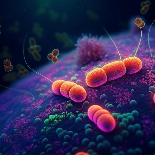
Explore how the benthic clamworm *Perinereis vancaurica* digests polystyrene foam debris, potentially transforming pollution into beneficial processes through intriguing gut microbiome interactions. This groundbreaking research by Sufang Zhao, Renju Liu, Shiwei Lv, Benjuan Zhang, Juan Wang, and Zongze Shao uncovers new opportunities for tackling plastic waste in marine ecosystems.
~3 min • Beginner • English
Introduction
The study addresses the fate of polystyrene (PS) foam debris in marine environments, particularly its fragmentation into microplastics and potential biodegradation within marine organisms. PS, widely used in packaging and aquaculture as expanded polystyrene (EPS), is prone to break into small fragments and contributes significantly to marine microplastics, which can be ingested by diverse marine fauna and accumulate through food webs. Despite widespread plastic pollution, the processes generating microplastics in situ and their biological degradation in marine systems remain poorly understood. During preliminary observations, EPS debris was found inhabited by burrowing clamworms that chewed EPS and excreted frass, suggesting possible biological fragmentation and microbial involvement. The research aims to determine the contribution of EPS-eating clamworms (Perinereis vancaurica) to microplastic formation and evaluate the role of their gut microbiome in PS digestion and degradation, expanding knowledge of marine benthic polychaetes in plastic biodegradation.
Literature Review
Prior work has primarily focused on terrestrial insect larvae degrading plastics. Mealworms (Tenebrio molitor) chew EPS, and PS-degrading bacteria such as Exiguobacterium sp. YT2 have been isolated from their guts; an Acinetobacter sp. was isolated from Tribolium castaneum larvae; Enterococcus has been implicated in PS degradation in Galleria mellonella and T. obscurus. Studies reported PS degradation products (styrene, dimers/trimers, and oxidized aromatics) in insect frass and tissues, and shifts in gut microbiota (e.g., Enterococcus, Enterobacteriaceae, Bacillus, Serratia, Pseudomonas, Lactococcus). Marine polychaete-mediated plastic degradation has not been previously documented. Related studies on Perinereis aibuhitensis indicate capacity to metabolize xenobiotics (e.g., PAHs) and harbor hydrocarbon-degrading bacteria (Pseudomonas, Vibrio). The current study contrasts with prior insect-based literature by focusing on a marine polychaete, indicating Acinetobacter and Ruegeria as key taxa rather than Enterococcus or Pseudomonas commonly seen in insect systems.
Methodology
Sampling and organism identification: EPS foam blocks with visible clamworm burrows were collected from the east coast of Xiamen Island, China (118.20°E, 24.49°N) in September 2021. Approximately twenty clamworms (5–12 cm) were found within EPS blocks. Morphological identification used stereomicroscopy. Molecular identification used mitochondrial 16S rRNA and COI gene amplification and neighbor-joining phylogenetic analysis (MEGA-7), confirming close relatedness to Perinereis vancaurica.
Frass and microplastic characterization: Excreted frass and EPS control were digested (10% KOH, 24 h, 50 °C; followed by H2O2, 24 h, 50 °C) and filtered. Particle sizes and morphology were assessed by stereomicroscopy (Leica M205A), light microscopy (Nikon 80i), and SEM (LEO-1530). EPS debris in the gut was visualized by stereomicroscopy and biofilms by fluorescence microscopy after LIVE/DEAD staining.
Chemical analyses in gut and frass: Clamworms were surface-cleaned; intestines were dissected. Intestine extracts (THF:methanol 2:1) and fresh frass extracts (THF; both re-dissolved in hexane) were analyzed by GC-MS (Agilent GC/MS-QP2010; DB-5MS UI column; helium carrier; oven 50 °C to 250 °C at 10 °C/min, 20 min hold; NIST14 library, ≥70% similarity). Micro-FTIR (Nicolet iN10) on gut EPS debris (670–4000 cm^-1, 16 scans, 8 cm^-1 resolution) quantified hydroxyl index (HI) and carbonyl index (CI) as ratios to methylene band area. FTIR (Nicolet iS50) compared frass vs EPS control. Gel permeation chromatography (Agilent 1260 Infinity II; THF 1 g L^-1, 0.7 mL/min) measured molecular weight distribution (MWD), Mw, Mn, and PDI (Mw/Mn). 1H NMR (Bruker Avance 400 MHz; CDCl3) analyzed frass chemistry.
Gut microbiome profiling: Two groups were used: nonplastic-fed (P0) animals from fishing shops (fed formulated animal feed) and EPS-fed (P1) animals maintained 15 days with EPS blocks in filtered seawater at room temperature. Intestines were dissected; bacterial 16S rRNA amplicons were sequenced (details in Supplementary Note S3). Frass-associated microbiota were also profiled from DNA extracted from frass in EPS tunnels.
Isolation of gut bacteria and PS-degradation assays: Intestinal homogenates were serially diluted and plated on Marine Broth 2216 and R2A agar. 28 isolates were identified by 16S rRNA gene sequencing and phylogeny (Supplementary Note S4). For PS utilization tests, isolates were inoculated into 50 mL carbon-free mineral medium (MMC) containing three sterile PS films (1 × 1 cm, thickness 0.19 mm; Sigma; pre-sterilized) as sole carbon source; incubated 30 days at 28 °C, 150 rpm, dark. Controls (CK) lacked bacteria. After incubation, PS films were washed (2% SDS, 4 h, 50 °C; rinse and dry). SEM examined biofilms and surface defects; fluorescence microscopy assessed viability.
Quantifying PS degradation by isolates: Weight loss was measured after 30 days (percentage = 100 × [(Initial − Final)/Initial]). FTIR-ATR (Thermo iS50; 500–4000 cm^-1, 4 cm^-1 resolution, 32 scans) evaluated functional group changes and carbonyl index. Water contact angle (Dataphysics OCA20) assessed hydrophilicity (2 µL droplets; average of triplicates). TGA (Mettler Toledo TGA 2; 30–550 °C, 10 °C/min, N2) evaluated thermal stability changes.
Metabolite profiling of bacterial degradation: Culture supernatants after 30 days were centrifuged, 0.22 µm filtered, extracted three times with chloroform:methanol (2:1), evaporated, re-dissolved in hexane, and analyzed via GC-MS as above. CK medium without bacteria was processed in parallel.
Key Findings
- EPS ingestion by Perinereis vancaurica resulted in fragmentation into microplastics and nanoplastics. Frass particle sizes ranged 0.3–1.3 mm with an average 0.6 ± 0.2 mm; nanoplastics 0.5–15 µm were also observed. SEM showed numerous cavities on frass microplastics vs smooth pristine EPS; bacterial biofilms were visible on microplastics within the gut.
- GC-MS of EPS-fed intestines detected long-chain fatty acids and esters and aromatic compounds: benzoic acid, 3,5-bis(1,1-dimethylethyl)-4-hydroxy; phenol, 2,6-bis(1,1-dimethylethyl)-4-ethyl; hexadecanoic acid, methyl ester; eicosanoic acid. Frass contained 4-methyl-2,6-di-tert-pentylphenol. These are structurally similar to intermediates reported from insect larvae feeding on PS, suggesting analogous degradation pathways.
- µFTIR of gut EPS debris showed increased hydroxyl and carbonyl signals vs control: HI = 11.9 and CI = 0.59 (vs control HI = 0.02; CI = 0.33), confirming oxidation and partial biodegradation in the intestine.
- FTIR of excreted frass showed new/enhanced bands near 1670 cm^-1 (aromatic-conjugated C=O) and 3310 cm^-1 (O–H), indicating oxidative modification.
- GPC showed depolymerization in frass: Mw decreased 2.1% (216.9 → 212.3 kDa), Mn decreased 12.5% (73.5 → 64.3 kDa), PDI decreased (3.3 → 2.95), and MWD shifted toward lower molecular weight fragments.
- 1H NMR of frass revealed new peaks in aliphatic (1–2 ppm) and aromatic (6–7 ppm) regions vs EPS control, indicating chemical structural changes.
- Gut microbiome shift with EPS diet: Family level—P0 dominated by Marinobacteraceae (49.51%) and Vibrionaceae (17.92%); P1 dominated by Moraxellaceae (80.98%), with Rhodobacteraceae (3.60%) and unclassified_c_Bacilli (3.40%). Genus level—P0: Marinobacter (49.5%), Vibrio (17.8%); P1: Acinetobacter (80.8%), Ruegeria (2.3%), unclassified_c_Bacilli (3.3%), Chryseobacterium (1.1%), Alcanivorax (1.2%), Vibrio (0.38%). Chi-square tests indicated significant shifts in Acinetobacter, Ruegeria, and Bacilli. Frass-associated microbiota in vitro were enriched in unclassified_f_Rhodobacteraceae (11.8%), Ruegeria (6.8%), Vibrio (4.6%), norank-Chloroplast (6.2%), and norank-Actinomarinales (5.2%).
- Ten of 28 gut isolates degraded PS in vitro: Acinetobacter (3), Brevibacterium (1), Gaetbulibacter (3), Ruegeria (2), Vibrio (1). Most effective strains were Acinetobacter johnsonii M1-3, Brevibacterium casei R3-1D, and Ruegeria arenilitoris M3-42 (MCCC 1A18346, 1A1836, 1A18356; 16S rRNA GenBank OP854692, OP854691, OP854690). SEM and live/dead staining showed dense viable biofilms and surface pits/cavities on PS films.
- PS film weight loss after 30 days: A. johnsonii M1-3, 3.7 ± 0.07%; B. casei R3-1D, 3.3 ± 0.06%; R. arenilitoris M3-42, 3.4 ± 0.18%; CK, 0.015 ± 0.003%.
- FTIR-ATR of films showed new carbonyl peaks at ~1725 cm^-1 and C–O at ~1060 cm^-1; carbonyl index increased markedly: M1-3 CI = 18.33; M3-42 CI = 24.17; R3-1D CI = 20.6; CK CI = 1.55. Water contact angle decreased from 93.9° ± 0.2 (CK) to 63.2° ± 0.4 (M1-3), 72.4° ± 0.2 (M3-42), 73.5° ± 0.2 (R3-1D), indicating increased hydrophilicity. TGA showed lower endothermic temperatures for treated films (413–414 °C) than control (421 °C), consistent with altered composition.
- GC-MS of culture supernatants revealed PS-derived aromatics and degradation products including ethylbenzene, o-xylene, benzaldehyde, 4-phenylbut-3-ene-1-yne, butylated hydroxytoluene, 3-butynylbenzene, pentanoic acid 2-phenylethyl ester, and a phthalate derivative, plus fatty acids/esters (e.g., butanoic acid; carbonic acid, dodecyl prop-1-en-2-yl ester; methoxyacetic acid, 2-tetradecyl ester) and hydrocarbons/alcohols. Detection supports the styrene oxide–phenylacetaldehyde pathway; butanoic acid in B. casei R3-1D was a novel observation among PS-degrading isolates.
Discussion
The findings demonstrate that a marine benthic polychaete, Perinereis vancaurica, actively chews EPS, leading to mechanical fragmentation and the production of micro- and nano-plastics. Concurrently, oxidative chemical modifications and partial depolymerization occur within the gut, as evidenced by increased hydroxyl and carbonyl indices, reduced Mn and Mw, and new NMR/FTIR features. The gut microbiome undergoes a marked shift under EPS feeding, becoming dominated by Acinetobacter (Moraxellaceae) and enriched in Ruegeria (Rhodobacteraceae) and Bacilli, indicating a specialized community potentially adapted to utilize or transform PS-derived substrates. Isolation of representative strains confirmed PS biodegradation in vitro by Acinetobacter johnsonii, Brevibacterium casei, and Ruegeria arenilitoris, causing measurable weight loss, oxidation (carbonyl formation), increased hydrophilicity, reduced thermal stability, and production of PS-derived monomers/oligomers and downstream metabolites. These results align with degradation pathways reported in insect larvae but highlight different dominant taxa in a marine host (Acinetobacter and Ruegeria vs Enterococcus/Pseudomonas commonly seen in insects), suggesting host- and environment-specific microbial consortia. Ecologically, clamworm activity can accelerate EPS fragmentation and introduce microplastics and associated chemicals into marine food webs, as clamworms are prey for higher trophic levels. The study underscores the dual role of mechanical and microbial processes in in situ microplastic generation and early-stage biodegradation in marine settings.
Conclusion
This work reports the first evidence of an EPS-chewing marine benthic polychaete (Perinereis vancaurica) and implicates its gut microbiome in PS biodegradation. EPS ingested by clamworms is fragmented into microplastics and exhibits oxidative modifications and partial depolymerization during gut passage. EPS feeding reshapes the gut microbiome, enriching Acinetobacter and Ruegeria, and isolates of Acinetobacter johnsonii, Brevibacterium casei, and Ruegeria arenilitoris degrade PS in vitro, causing 3.3–3.7% weight loss in 30 days and generating characteristic degradation intermediates. These findings highlight a marine biological pathway contributing to microplastic formation and early-stage biodegradation, with implications for plastic fate and ecological risk in marine ecosystems. Future work should identify specific degradative enzymes and pathways, quantify in situ degradation rates, optimize laboratory conditions to better replicate natural feeding, and assess the generality across Perinereis species and regions.
Limitations
- The extent of PS depolymerization and weight loss by both the host-associated process and isolated strains over 30 days was modest compared to some terrestrial insect systems, suggesting limited degradation under tested conditions.
- Clamworms likely did not rely on EPS as the sole carbon source in nature, potentially constraining biodegradation extent and complicating attribution between mechanical vs biochemical contributions.
- Specific enzymes and genetic pathways responsible for EPS degradation in the host and isolates were not identified.
- Laboratory EPS-feeding conditions may not fully replicate natural environmental variables; broader ecological relevance and rates remain to be quantified.
- The study focuses on one species and location; the ubiquity of this process across Perinereis species and regions requires further investigation.
Related Publications
Explore these studies to deepen your understanding of the subject.

