Medicine and Health
Polyphenol-rich diet mediates interplay between macrophage-neutrophil and gut microbiota to alleviate intestinal inflammation
D. Han, Y. Wu, et al.
Discover how dietary phenolic acids can combat intestinal inflammation by modifying gut microbiota and regulating macrophage activation! This groundbreaking research, conducted by Dandan Han and colleagues from China Agricultural University, reveals the unique interactions between individual phenolic acids and immune cells in the context of inflammatory bowel disease.
~3 min • Beginner • English
Introduction
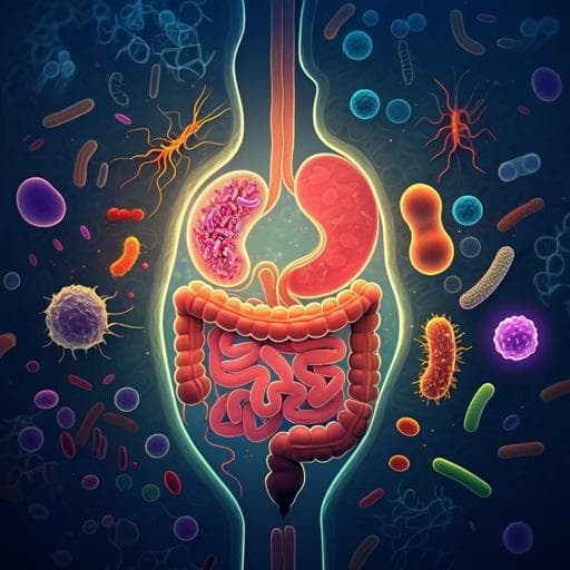
Inflammatory bowel disease (IBD) arises from dysregulated mucosal immune responses to resident microbiota, causing intestinal inflammation, tissue damage, and dysbiosis. Macrophages, owing to their plasticity, are central to intestinal homeostasis and are implicated in IBD pathogenesis, where skewing toward a proinflammatory M1 phenotype contributes to disease. Metabolic programming of macrophages (immunometabolism) regulates their phenotype and functions. Concurrently, gut microbiota maintains gastrointestinal integrity and immune homeostasis; alterations in composition and metabolites are linked to IBD, and microbiota is required for colitis initiation in DSS or IL-10-deficient mouse models. Therapeutic manipulation of the microbiota, including FMT from healthy donors, ameliorates colitis. Polyphenolic phenolic acids (chlorogenic, ferulic, caffeic, ellagic) improve gut health by modulating microbiota and immune responses, but how individual acids affect macrophage–microbiota–neutrophil interactions during colitis is unclear. The study investigates mechanisms by which distinct phenolic acids alleviate gut inflammation, focusing on macrophage and neutrophil roles and microbiota dependence.
Literature Review
Prior work implicates macrophages as key regulators in IBD, with M1/M2 polarization balance critical for resolution and homeostasis. Metabolic reprogramming, including glycolysis regulation, influences macrophage function and NLRP3 inflammasome activation. Gut microbiota composition and metabolites modulate intestinal integrity and immune responses; dysbiosis correlates with IBD severity, and microbiota is necessary for DSS colitis development. FMT from healthy or polyphenol-dosed donors can ameliorate colitis. Dietary polyphenols alter microbiota and reduce inflammation; specific phenolic acids (chlorogenic, ferulic, caffeic, ellagic) have been reported to affect gut health via microbiota shaping and immune modulation. Microbial metabolites of polyphenols (e.g., urolithins from ellagic acid) enhance gut barrier via Nrf2 and modulate immune cell IL-22 production. Neutrophils can drive or resolve inflammation; their extracellular traps (NETs) sustain inflammation in colitis, and inhibition of NETs attenuates disease. This literature motivates dissecting acid-specific mechanisms and the interplay among macrophages, neutrophils, and microbiota.
Methodology
- Animals: Male C57BL/6 mice housed under SPF conditions with standard chow and water. All procedures approved by China Agricultural University IACUC (AW51211202-1-3).
- DSS colitis: 3% DSS (36–50 kDa) in drinking water for 5 days, then water for 3 days. Disease assessed by body weight change, disease activity index (DAI), colon length, histology (H&E), myeloperoxidase (MPO) activity, cytokines.
- Microbiota depletion and FMT/metabolite transfer: Broad-spectrum antibiotics cocktail in water for 2 weeks (1 g/L streptomycin, 0.5 g/L ampicillin, 1 g/L gentamicin, 0.5 g/L vancomycin). Efficacy confirmed by bacterial DNA and total bacteria quantification. Fecal microbial suspensions prepared from donors (diluted in PBS with 15% glycerol), filtered through gauze and 0.22 µm filters; sterile fecal filtrate (SFF, metabolites) collected. ABX-treated mice conventionalized with fecal suspensions from SPF or phenolic acid–treated donors. For metabolite transfer, sterile fecal filtrates from caffeic acid (CA) or ellagic acid (EA) groups gavaged into DSS-treated recipients.
- Phenolic acid administration: In colitis experiments, mice received four phenolic acids (chlorogenic acid, caffeic acid, ferulic acid, ellagic acid) by oral gavage for 8 days; DSS started on day 3 for 5 days. For donor preparation in FMT studies, 7-week-old mice were gavaged once daily with CA or EA at 50 mg/kg in 100 µL PBS for two weeks.
- Macrophage depletion/transfer: Clodronate-loaded liposomes (200 µL i.p.) once every 3 days, three times prior to DSS; also on day 1 and day 4 during DSS or phenolic acid treatment. Some mice received 5×10^5 bone marrow–derived macrophages (BMDMs) i.p. on day 5 of DSS.
- Neutrophil depletion: Anti-Ly6G antibody (200 µg i.p., clone 1A8), or isotype IgG2a control, once every 3 days for three doses starting 2 days before DSS and on days 1 and 4 during DSS.
- NLRP3 inhibition: CY-09 administered to inhibit NLRP3 (dose/regimen as per referenced protocol); effects assessed on CGA responsiveness.
- Urolithin studies and IL-22 neutralization: DSS mice treated with urolithin A (UroA) or urolithin B (UroB) by gavage. For IL-22 requirement, anti-IL-22 mAb (50 µg i.p., clone 2G12A41) injected twice every 2 days; isotype control used.
- Cell culture: Caco2 epithelial cells, RAW264.7 macrophages, and BMDMs cultured in DMEM with 10% FBS. LPS stimulation used to induce inflammation. Co-culture: Caco2 on transwells with RAW264.7 in lower chamber; transepithelial electrical resistance (TEER) measured.
- RNA interference: siRNA knockdowns of Nlrp3 and Pkm2 in macrophages; knockdown verified by RT-qPCR.
- Readouts and assays: Flow cytometry for immune cell subsets (CD11b, CD11c, Gr-1, Ly6G, RORγt, IL-22, F4/80); ELISAs for cytokines (TNF-α, IL-1β, IL-6, IL-10, IL-18, IL-22), NLRP3, caspase-1 activity; glycolysis via Seahorse XF-96 extracellular acidification rate (ECAR); lactate assay; glucose transporter (Glut1) and glycolysis gene expression (Pkm2, Hk2, Pdk1, Ldha) by RT-qPCR; mitochondrial membrane potential and MitoSOX for ROS; NETs quantified by MPO-DNA complex ELISA. Histology scoring followed standard criteria. Statistics: Student’s t test or one-way ANOVA with Tukey post hoc; P<0.05 significant. Typical group sizes n=6–8 and three independent experiments.
Key Findings
- Interplay of microbiota and macrophages: Depletion of macrophages (clodronate) increased colitis severity by DAI without significant changes in weight loss or colon length; proinflammatory cytokines (TNF-α) increased and IL-10 decreased in colon; serum inflammatory factors elevated. Restoration with donor BMDMs reduced TNF-α and IL-1β and increased IL-10. Macrophage depletion altered microbiome composition, and FMT reduced F4/80+ macrophage infiltration in ABX+DSS mice, indicating crosstalk influences epithelial injury.
- Microbiota depletion: ABX induced ~4-log reduction in fecal bacteria and increased susceptibility to DSS (greater weight loss, DAI, and MPO activity). Barrier gene expression (Claudin-4, ZO-1) decreased; IL-10 secretion lower; mixed effects on TNF-α/IL-1β/IL-6 mRNA. FMT from SPF donors improved colon length, reduced MPO activity, and increased barrier gene expression; IL-6 decreased and IL-10 increased in colon tissue.
- Phenolic acids ameliorate colitis: Chlorogenic acid (CGA), caffeic acid (CA), ferulic acid (FA), and ellagic acid (EA) each reduced weight loss, DAI, colon shortening, histological inflammation, and systemic/colonic cytokines in DSS colitis. CGA and FA decreased colonic CD11b+CD11c+ M1 macrophages and downregulated M1 genes (Nos2, Cxcl9). In RAW264.7 cells, CGA and FA, but not CA or EA, reduced LPS-induced M1 gene expression (IL-1β, IL-6, TNF-α, Nos2, Cxcl9).
- CGA requires macrophages and NLRP3: In macrophage-depleted or NLRP3-inhibited (CY-09) mice, CGA failed to reduce colitis severity and systemic inflammation. CGA suppressed M1 cytokines and Nlrp3 mRNA in DSS colons under normal conditions but not with NLRP3 inhibition. Flow cytometry showed CGA did not reduce CD11b+CD11c+ M1 macrophages in NLRP3-deficient conditions. In Caco2–macrophage co-culture, CGA preserved TEER and reduced cytokines only with Nlrp3-WT macrophages, not Nlrp3-KD, indicating dependence on the macrophage–NLRP3 axis.
- CGA inhibits PKM2-dependent glycolysis: LPS increased glycolysis-related genes (Pkm2, Hk2, Pdk1, Ldha), Glut1, ECAR, and lactate in macrophages; CGA attenuated these changes without affecting viability. PKM2 knockdown reduced glycolytic gene expression, lactate, ECAR, and inflammasome activation (caspase-1, NLRP3) and IL-1β/IL-18 (not TNF-α). CGA’s inhibitory effects on glycolysis and associated inflammasome activation were lost in PKM2-deficient macrophages. CGA also mitigated mitochondrial dysfunction (membrane potential loss, MitoSOX), an effect reduced by PKM2 knockdown. Overall, CGA reduces M1 polarization by suppressing PKM2-dependent glycolysis and NLRP3 activation.
- FA acts via neutrophils and NETs: FA decreased neutrophil infiltration and neutrophil production of IL-17 and IL-22. Neutrophil depletion worsened colitis and abrogated FA’s anti-inflammatory effects (FA did not reduce cytokines in neutrophil-depleted mice). FA reduced NET formation (lower MPO–DNA complexes), indicating FA mitigates colitis partly by inhibiting NETs.
- CA and EA are microbiota-dependent: In ABX-treated mice, CA or EA failed to reduce colitis. FMT from CA- or EA-treated donors transferred protection to DSS recipients (reduced weight loss, DAI, colon shortening; lowered TNF-α, IL-6, IL-1β in serum and colon). Transfer of sterile fecal filtrates (metabolites) from EA-, but not CA-, treated donors reduced disease severity and decreased colonic Tnf-α, Il-1β, Il-6, and Il-22 mRNA, indicating metabolite-mediated effects for EA.
- Urolithin A (EA metabolite) protects via IL-22: UroA, but not UroB, reduced DSS colitis severity, cytokines (IL-1β, TNF-α, IL-6, IL-22), and improved barrier function (in vitro TEER and tight junction gene expression). IL-22 neutralization abolished UroA’s protective effects and its impact on ILC3s, demonstrating IL-22 dependence.
- Selected quantitative data: Clodronate group showed F4/80+ cells 57.65 ± 4.84% vs 28.23 ± 2.41% in controls; ABX produced ~4-log reduction in fecal bacteria. Group sizes typically n=6–8, with three independent experiments; statistical significance at P<0.05 or P<0.01.
Discussion
The study delineates distinct mechanisms by which individual dietary phenolic acids mitigate DSS-induced colitis through coordinated actions on immune cells and the microbiota. CGA’s benefit depends on macrophages and NLRP3 inflammasome activity and is mechanistically linked to suppression of PKM2-driven glycolysis, aligning immunometabolism with reduced M1 polarization and improved epithelial barrier function. FA’s effects are mediated in part by neutrophils, notably through inhibition of NET formation, underscoring neutrophil–macrophage cooperation in resolving inflammation. CA and EA require an intact microbiota to confer protection; microbiota from CA/EA-treated donors transfers anti-colitic effects. EA’s benefits are further attributable to microbial metabolites, particularly UroA, which enhances barrier integrity and reduces inflammation via IL-22-dependent pathways, likely acting on epithelial and innate lymphoid cells. Together, these findings integrate microbe–immune cell crosstalk and metabolic reprogramming, providing mechanistic insights and targets for modulating colitis severity.
Conclusion
Individual phenolic acids ameliorate intestinal inflammation through distinct, yet interconnected, pathways linking gut microbiota with macrophage and neutrophil functions. Chlorogenic acid alleviates colitis by inhibiting PKM2-dependent glycolysis in macrophages and suppressing NLRP3 activation, thereby reducing M1 polarization and enhancing barrier function. Ferulic acid reduces disease at least partly by inhibiting neutrophil extracellular trap formation, indicating a neutrophil-dependent mechanism. Caffeic acid and ellagic acid require the gut microbiota to confer protection; fecal microbiota from CA/EA-treated donors transfers benefits, and metabolites derived from EA (not CA), particularly urolithin A, mitigate colitis by promoting barrier integrity in an IL-22-dependent manner. These results suggest phenolic acids as promising candidates for managing intestinal inflammation by targeting microbiota–immune cell–metabolism axes. Future work should validate these effects in human IBD and define molecular targets (e.g., receptors and downstream signaling) to optimize therapeutic strategies.
Limitations
Findings are based on murine DSS colitis models and may not directly translate to human IBD. The specific receptors and downstream signaling pathways through which phenolic acids (and their metabolites) act on immune and epithelial cells remain undefined. Not all dosing regimens were standardized across acids in all experiments, and some mechanistic dependencies (e.g., precise contributions of other immune subsets) were not fully dissected. The apparent discrepancy in macrophage depletion markers and potential off-target effects of depletion/inhibition strategies (clodronate liposomes, CY-09) may influence interpretations. Clinical efficacy, safety, and microbiota variability in humans require further investigation.
Related Publications
Explore these studies to deepen your understanding of the subject.


