BiologyCommunications Physics
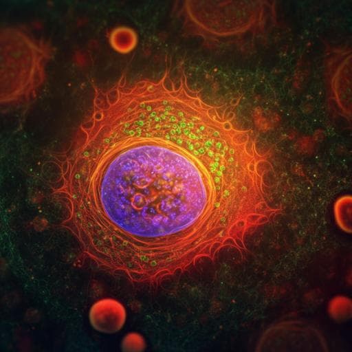
Optogenetic control of migration of contractile cells predicted by an active gel model
O. M. Drozdowski, F. Ziebert, et al.
This exciting research by Oliver M. Drozdowski, Falko Ziebert, and Ulrich S. Schwarz explores how optogenetics can control actin-driven cell migration by manipulating myosin contractility. The study reveals a bistability between sessile and motile states that opens new possibilities for understanding cellular behavior in response to light, with implications consistent with real-world neutrophil experiments.
Related Publications
Explore these studies to deepen your understanding
Adjacent work that informs or extends this paper's methodology and findings.

Physics
Active-feedback quantum control of an integrated low-frequency mechanical resonator
J. Guo, J. Chang, et al.
Biology
Dopamine control of social novelty preference is constrained by an interpeduncular-tegmentum circuit
S. Molas, T. G. Freels, et al.

Psychology
Dopamine control of social novelty preference is constrained by an interpeduncular-tegmentum circuit
S. Molas, T. G. Freels, et al.

Social Work
The influence of enterprise dormitories on the urban integration of migrant workers in China: an exploration of two distinct migration stages of individual and family migration and the differences between them
W. Wei and L. Zhang


