Biology
Open Exercise Promotes Tissue Regeneration: Mechanisms Involved and Therapeutic Scope
C. Liu, X. Wu, et al.
Discover how exercise can invigorate your body's regenerative abilities! This insightful review by Chang Liu, Xinying Wu, Gururaja Vulugundam, Priyanka Gokulnath, Guoping Li, and Junjie Xiao dives into the profound impact of physical activity on tissue regeneration in skeletal muscle, the nervous system, and the vascular system, while unraveling the key molecular mechanisms at play.
~3 min • Beginner • English
Introduction
Exercise exerts protective and regenerative effects in multiple tissues, including cardiac, neural, and skeletal muscle. Diverse exercise modalities (e.g., swim training, treadmill running, voluntary wheel running) produce specific cellular responses and regenerative outcomes that vary with intensity and duration. Because many individuals cannot meet exercise intensity/duration thresholds, elucidating molecular mechanisms by which exercise promotes regeneration is crucial to enable translational therapies. This review aims to summarize effects of exercise on tissue regeneration—focusing on stem/progenitor cell activation in muscle, brain, and vasculature—describe underlying molecular mechanisms (growth factor signaling, oxidative stress and ROS, metabolic reprogramming, and non-coding RNAs), and discuss therapeutic strategies that pharmacologically target these pathways to mimic exercise-induced regeneration.
Literature Review
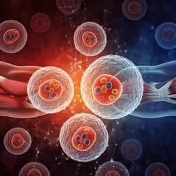
Muscle regeneration: Adult skeletal muscle relies on quiescent Pax7+ muscle satellite cells (MSCs) for repair and growth. Exercise activates MSCs to proliferate, differentiate, and self-renew, contributing to myonuclear accretion context-dependently. Aging disrupts MSC niche cues (e.g., altered ECM, Smoc2 changes) and can induce fibrogenic conversion, reducing myogenicity. Exercise counteracts atrophy and promotes MSC activation and self-renewal via AKT, MAPK, and metabolic reprogramming, with age-specific differences (e.g., AKT pathway activation and cyclin D1 restoration in aged mice; MAPK signaling and mitochondrial repression to preserve stemness in young mice). Cross-talk with other cell types (e.g., AMPK-driven FAP senescence) supports regenerative inflammation and repair. Neural regeneration: Adult neurogenesis occurs chiefly in the hippocampal dentate gyrus (DG) and is robustly enhanced by voluntary running, increasing proliferation and neuronal differentiation of neural progenitor cells (NPCs). Exercise improves cognitive deficits in disease models (e.g., Alzheimer’s), increases oligodendrocyte precursor cells in hypoperfusion models, and enhances synaptogenesis and plasticity. Mechanisms include VEGF, IGF1 (via IGF1–RIT1–Akt–Sox2), GH, serotonin, BDNF (including BBB-crossing influences via myokines like cathepsin B and metabolites like lactate), PGC‑1α/FNDC5 signaling, ROS dynamics (transient ROS surge recruits hiROS NPCs), Notch signaling in progenitor survival/cell-cycle exit, RGS6 in maturation of newborn neurons, and platelet-derived PF4. Cardiac regeneration: While adult hearts lack bona fide cardiac stem cells, exercise promotes endogenous cardiomyocyte proliferation and repair after injuries (MI, IRI, pressure overload, doxorubicin cardiomyopathy). Mechanisms include IGF–PI3K–Akt signaling with downregulation of C/EBPβ and upregulation of CITED4, and regulation by non-coding RNAs: miR‑222 and miR‑17‑3p facilitate proliferation and cardioprotection; ADAR2–miR‑34a–cyclin D1 axis supports regeneration; lncRNAs (LncCPhar, lncExACT1) modulate cardiomyocyte proliferation and physiological hypertrophy. Angiogenesis and lymphangiogenesis: Exercise enhances angiogenesis (VEGF pathway) and lymphangiogenesis (e.g., cardiac VEGFR3 upregulation), supporting regeneration and physiological hypertrophy. Exercise increases circulating endothelial progenitor cells (EPCs), improves EPC homing to ischemic tissues, and augments EPC-derived exosome function, with documented benefits across aging and disease models. Collectively, studies demonstrate that exercise-driven stem/progenitor activation and vascular remodeling are central to regeneration across organ systems, mediated by conserved signaling and regulatory RNA networks.
Methodology
This is a narrative review. The authors identified literature primarily via PubMed searches using keywords in combinations such as: exercise, tissue regeneration, muscle stem cells, neural stem cells, cardiomyocytes, and regenerative therapy. Only English-language articles were included; abstracts and meeting reports were excluded. The review integrates findings from animal and human studies across exercise modalities (e.g., rodent swim training, treadmill running, voluntary wheel running) and durations to examine common and tissue-specific mechanisms underlying exercise-induced regeneration. Mechanistic evidence from genetic, molecular, and pharmacological studies was synthesized to outline therapeutic targets that mimic exercise benefits.
Key Findings
- Exercise activates stem/progenitor cells to promote regeneration: MSCs in skeletal muscle, NPCs in the hippocampal DG, endogenous cardiomyocytes in the heart, and EPCs for vascular repair. Age modifies responses; exercise rejuvenates aged MSCs and restores NSC numbers in aged brains.
- Muscle: Exercise counters atrophy and enhances MSC proliferation/self-renewal via AKT and MAPK pathways and metabolic reprogramming (reduced mitochondrial respiration to preserve stemness). In aged mice, long-term exercise activates AKT and restores cyclin D1; endurance exercise promotes MSC self-renewal and inhibits differentiation. Exercise-induced AMPK activation promotes FAP senescence, fostering regenerative inflammation.
- Brain: Voluntary running increases hippocampal NPC proliferation and neurogenesis; improves cognition in Alzheimer’s models; increases OPCs under hypoperfusion. Key mediators: VEGF, IGF1 (via RIT1–Akt–Sox2), GH, serotonin, BDNF (including muscle-brain crosstalk via cathepsin B, lactate; PGC‑1α/FNDC5). ROS dynamics recruit hiROS NPCs to proliferation; Notch1 supports survival/cell-cycle exit; RGS6 is required for running-enhanced neurogenesis; PF4 from activated platelets promotes neurogenesis.
- Heart: Exercise induces cardiomyocyte proliferation and cardioprotection post-injury (MI, IRI, pressure overload, doxorubicin). Mechanisms: IGF–PI3K–Akt axis downregulates C/EBPβ and upregulates CITED4; non-coding RNAs including miR‑222, miR‑17‑3p, ADAR2–miR‑34a–cyclin D1, LncCPhar, and lncExACT1 regulate proliferation and physiological hypertrophy.
- Vascular/lymphatic: Exercise promotes angiogenesis (VEGF pathway) and cardiac lymphangiogenesis via VEGFR3, supporting physiological hypertrophy and cardiomyocyte proliferation. Exercise elevates circulating EPCs, enhances EPC homing, improves arterial elasticity in aged men, and boosts EPC-derived exosome function.
- Therapeutic scope: Pharmacologically targeting IGF1 (protein, small molecules like BGP‑15, AAV gene therapy) and PI3K–Akt (PTEN inhibitors; PI3K gene therapy) can mimic exercise benefits in muscle, brain, and heart. Exercise-regulated miRNAs (e.g., miR‑23a/27a, miR‑29b suppression, miR‑135a, miR‑17‑3p, miR‑222) show therapeutic potential in muscle atrophy, neurogenesis, and cardioprotection.
Discussion
The collected evidence supports the hypothesis that exercise promotes tissue regeneration primarily through activation and modulation of resident stem/progenitor cells and pro-regenerative signaling networks. This activation occurs across multiple organs and is mediated by conserved pathways (IGF–PI3K–Akt, MAPK, Notch), metabolic and redox cues (ROS dynamics, mitochondrial regulation), and non-coding RNA programs (miRNAs, lncRNAs). These mechanisms translate into functional benefits in disease and aging contexts—enhanced muscle repair and hypertrophy, improved cognitive function and neurogenesis, cardiomyocyte proliferation and post-injury recovery, and augmented angiogenesis/lymphangiogenesis. Importantly, the review underscores that age, exercise type, and duration influence outcomes, suggesting tailored approaches for maximal benefit. The therapeutic translation leverages exercise-identified targets to design pharmacologic and gene therapies that emulate exercise-induced regeneration, addressing populations unable to exercise adequately. However, heterogeneity of models and incomplete mechanistic mapping necessitate further work to pinpoint causal targets, optimize delivery, and ensure cell type-specific effects.
Conclusion
Exercise exerts robust pro-regenerative effects in skeletal muscle, nervous, cardiovascular, and vascular systems through activation of stem/progenitor cells and modulation of key signaling, metabolic, oxidative, and non-coding RNA pathways. Targeting pivotal mediators such as IGF1, PI3K–Akt, and exercise-regulated miRNAs can mimic these benefits and represents a promising avenue for regenerative therapies. Future research should: (1) account for modulators like age to refine phenotypic assessments; (2) explore combination strategies where exercise acts synergistically with exogenous pro-regenerative factors; (3) ensure precise, cell type-specific targeting to avoid opposing effects; and (4) deepen mechanistic understanding to expand the therapeutic repertoire for degenerative diseases.
Limitations
- Variability by age, exercise type, intensity, and duration can alter regenerative outcomes and mechanisms, complicating generalization.
- Exercise may be insufficient alone in certain contexts and may require co-administration of pro-regenerative factors.
- Exercise-regulated molecules can have cell type-specific and potentially opposing effects, necessitating precise targeting in translational applications.
- Mechanisms, while outlined, remain incompletely defined; causal nodes and direct targets in many pathways (e.g., specific non-coding RNAs and their networks) need clarification.
- Therapeutic translation of IGF1/PI3K signaling faces challenges including conflicting clinical outcomes and risks such as tumorigenesis or off-target effects; optimized delivery, dosing, and tissue targeting are required.
- As a narrative review limited to English-language, peer-reviewed articles (excluding abstracts/reports), selection bias and publication bias may influence the synthesis.
Related Publications
Explore these studies to deepen your understanding of the subject.


