Medicine and HealthNature Communications
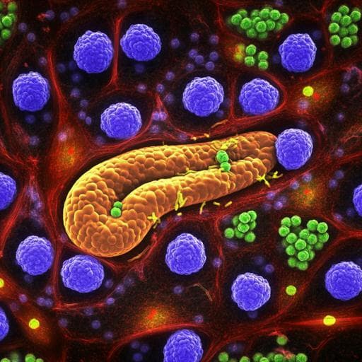
NEK2 inhibition triggers anti-pancreatic cancer immunity by targeting PD-L1
X. Zhang, X. Huang, et al.
This groundbreaking study reveals how NEK2 kinase phosphorylates PD-L1, hindering the impact of PD-L1-targeted immunotherapy in pancreatic cancer. The authors discovered that inhibiting NEK2 improves lymphocyte infiltration and enhances the immune response, offering a novel strategy for advancing pancreatic cancer treatment.
Related Publications
Explore these studies to deepen your understanding
Adjacent work that informs or extends this paper's methodology and findings.
Medicine and Health
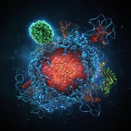
Hsc70 promotes anti-tumor immunity by targeting PD-L1 for lysosomal degradation
X. Xu, T. Xie, et al.
Medicine and Health
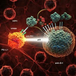
Targeting USP2 regulation of VPRBP-mediated degradation of p53 and PD-L1 for cancer therapy
J. Yi, O. Tavana, et al.

Medicine and Health
Radiogenomics for predicting p53 status, PD-L1 expression, and prognosis with machine learning in pancreatic cancer
Y. Iwatate, I. Hoshino, et al.
Medicine and Health
Efficacy and safety of serplulimab plus nab-paclitaxel in previously treated patients with PD-L1-positive advanced cervical cancer: a phase II, single-arm study
J. An, X. Li, et al.


