Medicine and HealthNutrition and Diabetes
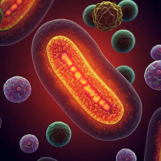
N-acetylcysteine supplementation did not reverse mitochondrial oxidative stress, apoptosis, and inflammation in the salivary glands of hyperglycemic rats
Z. Anna, K. Joanna, et al.
This study by Zalewska Anna and colleagues explores whether N-acetylcysteine supplementation can counteract harmful effects of a high-fat diet on salivary gland function in hyperglycemic rats. Discover how NAC influences mitochondrial activity and inflammatory markers, and what implications this may have for oxidative stress management.
Related Publications
Explore these studies to deepen your understanding
Adjacent work that informs or extends this paper's methodology and findings.

Medicine and Health
Inflammation and Oxidative Stress in Frailty and Metabolic Syndromes-Two Sides of the Same Coin
D. S and M. M

Medicine and Health
Mitochondrial Oxidative Stress Is the General Reason for Apoptosis Induced by Different-Valence Heavy Metals in Cells and Mitochondria
S. M. Korotkov
Veterinary Science
Consumption of a high energy density diet triggers microbiota dysbiosis, hepatic lipidosis, and microglia activation in the nucleus of the solitary tract in rats
D. M. Minaya, A. Turlej, et al.

Humanities
A "pre-traumatic stress syndrome": trauma and war in Elizabeth Bowen's *The Last September* and *The Heat of the Day*
Q. He


