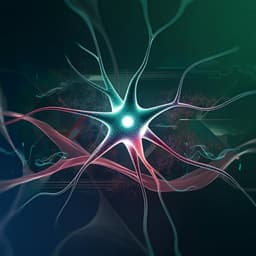
N-acetylaspartate (NAA) is a prominent metabolite in the central nervous system (CNS), synthesized by NAT8L and broken down by aspartoacylase (ASPA). While primarily found in neurons, NAA can be released extracellularly and taken up by glial cells. Elevated blood NAA levels are observed in neurodegenerative diseases like ALS. Although ASPA is expressed in various peripheral tissues including skeletal muscle, its role outside the CNS is understudied. Skeletal muscle comprises diverse myofibers categorized by contraction speed and energy source: type I (slow-twitch, oxidative), type IIA (intermediate), and type IIB/X (fast-twitch, glycolytic). The relative abundance of these fiber types is influenced by factors like training, age, and disease. Notably, a glycolytic-to-oxidative fiber-type switch is observed in ALS, with muscles exhibiting increased lipid oxidative metabolism and a shift from type IIB to type IIA myosin heavy chain (MHC) isoforms. Given the metabolic alterations in ALS muscle, the increase in serum NAA levels in ALS patients, and the potential local NAA increase due to motor neuron degeneration, this study aimed to investigate NAA's contribution to the rewiring of muscle cell metabolism.
Literature Review
Previous research has established NAA's role in the CNS, particularly its synthesis in neurons and catabolism in glial cells. Studies have linked elevated NAA levels in cerebrospinal fluid and blood to neurodegenerative diseases. However, the function of the NAA pathway in peripheral tissues, especially skeletal muscle, remains largely unexplored. Existing literature highlights the metabolic heterogeneity of skeletal muscle fibers and the impact of various factors on fiber type composition. Studies on ALS have demonstrated a metabolic reprogramming in skeletal muscle, involving a shift towards oxidative metabolism and a change in MHC isoform expression. This study builds upon these existing findings by directly investigating the impact of NAA on skeletal muscle metabolism and its potential role in the adaptation of muscle cells to disease.
Methodology
This study utilized differentiated C2C12 myotubes as an in vitro model of skeletal muscle. NAA was administered after 5 days of differentiation for 48 hours. Various assays were performed to assess the effects of NAA on different aspects of myotube metabolism and phenotype. Acetyl-lysine levels and lipid droplet (LD) content were analyzed using Western blotting and Oil Red-O staining. ATP production was measured using a colorimetric assay, and extracellular lactate levels were determined to assess glycolytic rate. Mitochondrial biogenesis and activity were evaluated through Western blotting and assays of mitochondrial complex I activity. Myofiber identity was assessed by analyzing the expression levels of different MHC isoforms using RT-qPCR. Myotube diameter was measured to assess phenotypic changes. The roles of protein synthesis and degradation pathways were investigated by analyzing the phosphorylation levels of mTOR-S6K pathway components, levels of ubiquitinated proteins, and the activation of autophagy. To determine the role of NAA catabolism, ASPA knockout (KO) C2C12 cells were generated using CRISPR/Cas9 technology. The response of NAA-treated myotubes to atrophic stimuli (dexamethasone, TNFα, and cisplatin) was also evaluated. Finally, the study utilized the SOD1-G93A ALS mouse model to investigate ASPA expression in muscles undergoing a glycolytic-to-oxidative switch. NAA levels were measured in serum, spinal cord, and brown adipose tissue (BAT) using HPLC.
Key Findings
NAA treatment significantly increased acetyl-lysine levels and enhanced lipid turnover in C2C12 myotubes, promoting both lipogenesis and lipolysis. This effect was dependent on ASPA-mediated NAA catabolism, as ASPA KO cells showed no response to NAA. NAA treatment resulted in increased ATP production, primarily through oxidative phosphorylation fueled by fatty acid oxidation. Mitochondrial biogenesis and activity were also significantly enhanced, as evidenced by increased levels of PGC1α, NRF1, ACO2, and TFAM. Consistently, NAA treatment induced a decrease in extracellular lactate, indicating a reduction in glycolytic rate. NAA treatment promoted a phenotypic switch towards oxidative myofibers, characterized by decreased MHC IIb and increased MHC IIa and MHC 7b, along with increased myoglobin expression. NAA treatment also led to a reduction in myotube diameter, likely due to increased protein turnover, involving both enhanced protein synthesis and increased proteasomal and autophagic degradation. Importantly, NAA-treated myotubes demonstrated increased resistance to atrophic stimuli (dexamethasone, TNFα, and cisplatin). In the SOD1-G93A ALS mouse model, increased ASPA levels were observed in muscles undergoing a glycolytic-to-oxidative switch, correlating with increased ATGL and HSL lipase levels. Serum NAA levels were elevated in ALS mice, accompanied by decreased NAA in the spinal cord and increased NAA in BAT, suggesting potential sources of NAA influencing muscle metabolism.
Discussion
This study reveals a novel role for NAA catabolism in regulating skeletal muscle metabolic identity. The findings demonstrate that NAA promotes a metabolic switch from glycolysis to oxidative phosphorylation, primarily through the activation of lipid turnover. This metabolic shift is coupled with phenotypic changes characteristic of oxidative myofibers and increased resistance to atrophy. The dependence of these effects on ASPA-mediated NAA catabolism highlights the importance of this metabolic pathway in muscle adaptation. The observed upregulation of ASPA in ALS mouse models undergoing a similar metabolic switch suggests that NAA could function as a compensatory mechanism, promoting a more resistant oxidative phenotype in diseased muscle. The study also explores potential sources of NAA influencing muscle metabolism, including neuronal degeneration and BAT hyperactivation in ALS. This study provides a valuable insight into the intricate interplay between NAA metabolism and skeletal muscle adaptation in both physiological and pathological contexts.
Conclusion
This study demonstrates that NAA catabolism plays a critical role in regulating skeletal muscle metabolism and phenotype, promoting a shift towards oxidative fibers and enhancing resistance to atrophy. The findings suggest that NAA could serve as a protective mechanism in conditions like ALS, where a metabolic switch towards oxidative metabolism is observed. Future studies could focus on further elucidating the molecular mechanisms underlying NAA's effects on muscle metabolism and exploring the therapeutic potential of targeting the NAA pathway for treating muscle atrophy.
Limitations
The study primarily utilizes an in vitro model (C2C12 myotubes), which may not fully capture the complexity of in vivo muscle physiology. While the SOD1-G93A ALS mouse model provides valuable in vivo data, it is still a model and may not completely reflect the human condition. Further investigation is needed to fully understand the contribution of different sources of NAA to muscle metabolism in vivo, including local production within the muscle tissue. The specific molecular mechanisms through which NAA influences protein turnover and resistance to atrophy warrant further investigation.
Related Publications
Explore these studies to deepen your understanding of the subject.