
BiologyNature Methods
Multielement Z-tag imaging by X-ray fluorescence microscopy for next-generation multiplex imaging
M. Strotton, T. Hosogane, et al.
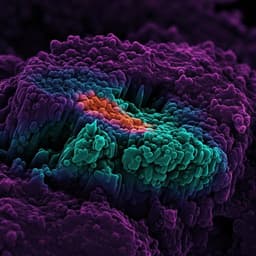
Discover the groundbreaking multielement Z-tag X-ray fluorescence (MEZ-XRF) bioimaging method developed by Merrick Strotton, Tsuyoshi Hosogane, Marco di Michiel, Holger Moch, Zsuzsanna Varga, and Bernd Bodenmiller. This innovative technique allows for rapid, high-resolution imaging of subcellular features, marking a significant advancement in multiomic tissue analysis.
Related Publications
Explore these studies to deepen your understanding
Adjacent work that informs or extends this paper's methodology and findings.

Medicine and Health
COVID-19 Prognosis from Chest X-ray Images by using Deep Learning Approaches: A Next Generation Diagnostic Tool
M. Pal, S. Parij, et al.
Engineering and Technology
Bottom-up construction of low-dimensional perovskite thick films for high-performance X-ray detection and imaging
S. Dong, Z. Fan, et al.

Engineering and Technology
Enhanced detection of threat materials by dark-field x-ray imaging combined with deep neural networks
T. Partridge, A. Astolfo, et al.

Physics
Resolution-enhanced X-ray fluorescence microscopy via deep residual networks
L. Wu, S. Bak, et al.


