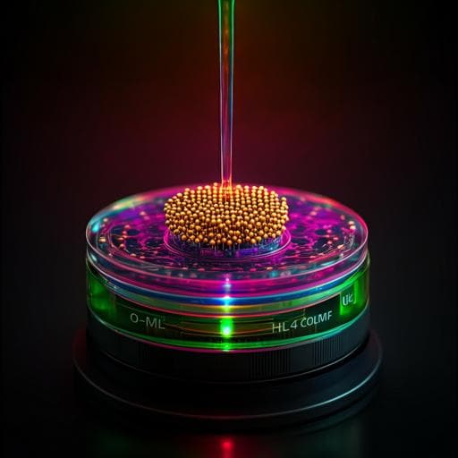
Monolithic integration of nanorod arrays on microfluidic chips for fast and sensitive one-step immunoassays
Y. Wang, J. Zhao, et al.
Explore these studies to deepen your understanding
Adjacent work that informs or extends this paper's methodology and findings.
Controlled on-chip fabrication of large-scale perovskite single crystal arrays for high-performance laser and photodetector integration
Z. Xu, X. Han, et al.
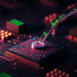
Highly stable integration of graphene Hall sensors on a microfluidic platform for magnetic sensing in whole blood
N. Shah, V. Iyer, et al.

Neuroimaging the effects of smartphone (over-)use on brain function and structure-a review on the current state of MRI-based findings and a roadmap for future research
C. Montag and B. Becker

Neuroimaging the effects of smartphone (over-)use on brain function and structure—a review on the current state of MRI-based findings and a roadmap for future research
C. Montag and B. Becker


