
Medicine and Health
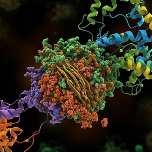
Molecular insights into receptor binding energetics and neutralization of SARS-CoV-2 variants
M. Koehler, A. Ray, et al.
This cutting-edge study reveals how SARS-CoV-2 mutations, particularly N501Y and E484Q, modify receptor binding and antibody neutralization. By employing atomic force microscopy and molecular dynamics, the authors delve into the stability of the RBD-ACE2 complex, offering crucial insights into the variant's implications on immunity. This research was conducted by Melanie Koehler, Ankita Ray, Rodrigo A. Moreira, Blinera Juniku, Adolfo B. Poma, and David Alsteens.
~3 min • Beginner • English
Introduction
SARS-CoV-2 continues to evolve with variants of concern (VoCs) that display increased transmissibility and reduced susceptibility to infection- and vaccine-induced immunity. Entry is a key step in the SARS-CoV-2 life cycle, mediated by the spike glycoprotein whose receptor-binding domain (RBD) engages the human ACE2 receptor. Mutations in the spike, including those near the fusion region (e.g., D614G) and within the receptor-binding motif (RBM), can alter ACE2 binding and antibody recognition. The RBD is the primary target of neutralizing antibodies induced by infection and vaccines; however, antigenic variation in residues such as E484 and N501 has led to therapeutic antibodies losing efficacy. The research question addressed here is how RBD mutations in VoCs (Alpha, Beta, Gamma, Kappa; with some comparisons to Delta) quantitatively impact the kinetic, thermodynamic, and structural parameters of RBD–ACE2 binding, and how these changes influence neutralization by monoclonal antibodies. The study combines single-molecule force spectroscopy using atomic force microscopy (AFM) with all-atom molecular dynamics (MD) simulations to dissect binding energetics and interface remodeling at atomistic detail.
Literature Review
Background work established that SARS-CoV-2 spike engages ACE2 with high affinity and that mutations (e.g., D614G) can increase infectivity by modulating spike conformational states. The RBD RBM tolerates mutations that can enhance ACE2 binding (e.g., N501Y) while compromising certain monoclonal antibodies (e.g., E484K leading to resistance and impacting bamlanivimab efficacy). Prior AFM and biolayer interferometry studies reported high-affinity WT RBD–ACE2 interactions (~10 nM) and multivalent interfacial contacts. Structural studies and computational analyses also highlighted residues 417, 484, and 501 as critical determinants of ACE2 affinity and antibody recognition.
Methodology
Experimental: Force–distance (FD) AFM was used to quantify single-molecule interactions between RBD variants and ACE2. ACE2 was covalently immobilized on gold-coated surfaces via EDC/NHS chemistry. AFM tips were functionalized with VoC RBD proteins through PEG linkers. Tips were approached/retracted cyclically to generate force-time/force-distance curves. Specific unbinding events were identified by rupture distances >12 nm (PEG extension). Binding frequencies (BF) were determined at defined contact times and approach/retract speeds. Dynamic force spectroscopy (DFS) was performed across multiple loading rates (LR) and hold times. Single-bond rupture forces versus LR were analyzed using the Bell–Evans model to extract kinetic off-rates (koff). BF versus contact time data were fitted to pseudo-first-order association kinetics to estimate kon and derive Kd = koff/kon, accounting for effective concentration within an accessible volume defined by linker and protein dimensions. Controls included PEG-only tips and surfaces lacking ACE2 to confirm specificity. Antibody inhibition assays: Binding between ACE2 and RBD (WT, Alpha, Beta, Gamma, Kappa) was measured before and after incubation with monoclonal anti-RBD antibodies (B-K45 and B-R41) or an IgG isotype control at 1, 10, and 50 µg mL−1. Reductions in BF were used to estimate inhibition efficacy and approximate IC50. Computational: All-atom MD simulations (GROMACS, CHARMM36m, TIP3P water) were performed on solvated RBD–ACE2 complexes for WT and VoCs (Alpha, Beta, Gamma, Kappa). Systems were energy-minimized, equilibrated (NVT then NPT), and simulated in production runs (multiple 250 ns replicas; cumulative tens of microseconds). Analyses included Cα RMSD/RMSF for stability, interfacial non-bonded energy decomposition (Coulomb and Lennard–Jones) between residue pairs across the interface, and contact frequency maps. Energetic contributions of key mutated residues (417, 452, 478, 484, 501) and conserved residues near the interface were quantified.
Key Findings
- AFM binding propensity: Specific binding frequencies followed Alpha-RBD > Beta-RBD > Gamma-RBD > Kappa-RBD > WT-RBD, indicating facilitated ACE2 engagement by VoCs relative to WT.
- Single-bond kinetics and stability: Bell–Evans analysis showed reduced koff (higher stability) for VoCs relative to WT (WT koff ≈ 0.011 ± 0.006 s−1; order of decreasing koff: WT > Alpha > Beta > Kappa > Gamma). Estimated lifetimes under the tested conditions increased across variants, with approximate values: Beta ~125 ms, Kappa ~165 ms, Gamma ~500 ms; Kappa and Gamma formed roughly twofold and fivefold more stable complexes than WT, respectively.
- Affinity (Kd) from BF vs time: All RBD–ACE2 interactions were high affinity. Reported equilibrium dissociation constants (ascending affinity; lower is tighter): Gamma 21 ± 16 nM < Kappa 31 nM < Beta 48.9 ± 1 nM < Alpha 121.8 ± 1 nM < Delta 134.1 nM. Gamma and Kappa exhibited lower Kd (stronger binding) than WT (WT value referenced in Supplementary data).
- MD energetics and contacts: The WT interfacial potential energy (sum of Coulomb + LJ) was ~−190.22 kJ/mol. The Kappa complex showed a different energetic balance (reported total energy ~−152.1 kJ/mol) yet formed a stabilized interface via redistributed contacts. Alpha, Beta, and Gamma exhibited gains in Lennard–Jones energies (~10 kJ/mol more favorable than WT), consistent with additional van der Waals/hydrogen bond contacts, especially around RBD residues 484 and 501.
- Mutation-specific effects: N501Y (present in Alpha, Beta, Gamma) had the strongest impact, adding contacts (e.g., hydrophobic Y501–Y41 in ACE2) and ~10 kJ/mol LJ energy gain. E484K/E484Q altered charge and salt-bridge networks involving ACE2 K31, modulating local electrostatics and stability. K417N/T substitutions weakened or abolished the WT K417–D30 salt bridge. Kappa showed high-frequency contacts with ACE2 K353 via RBD G496 and Q498, stabilizing the ACE2 β-hairpin region and broadening the interfacial footprint.
- Antibody inhibition: mAb B-K45, raised against WT-RBD, inhibited ACE2 binding for WT and all VoCs with ~50% reduction at 1 µg mL−1 (IC50 in low µg mL−1 range). mAb B-R41 effectively inhibited WT and Alpha but lost most neutralization against Beta, Gamma, and Kappa (variants sharing mutation at E484), showing only ~20–30% BF reduction.
Discussion
Combining single-molecule AFM and atomistic MD establishes that multiple SARS-CoV-2 VoCs enhance the stability and affinity of the RBD–ACE2 complex relative to WT, offering a mechanistic basis for increased transmissibility through improved receptor engagement. Enhanced stability arises from distinct mutation-dependent mechanisms: N501Y creates a binding hotspot with added hydrophobic contacts, while mutations at E484 and K417 rewire electrostatic networks and salt bridges. Kappa achieves a more balanced stabilization by distributing contacts across the interface, notably engaging ACE2 K353 via RBD G496 and Q498, thereby reinforcing the ACE2 β-hairpin anchoring. These structural and energetic adjustments extend bond lifetimes under force and reduce koff, aligning with higher BFs and lower Kd values for several VoCs. Neutralization assays reveal that while some antibodies (B-K45) retain broad efficacy across variants, others (B-R41) are compromised by mutations at E484, consistent with known immune escape at this position. Overall, the findings link specific mutational changes to altered interfacial energetics and kinetics, illuminating how SARS-CoV-2 evolves to maintain or improve receptor binding while evading subsets of antibodies.
Conclusion
This study provides quantitative kinetic, thermodynamic, and structural insights into how RBD mutations in SARS-CoV-2 VoCs modulate ACE2 binding. AFM revealed higher binding frequencies, reduced koff, and high-affinity interactions for VoCs, with Gamma and Kappa showing the strongest affinities among those tested. MD pinpointed residue-level energetic contributions, highlighting N501Y as a major stabilizer and E484/K417 substitutions as key modulators of electrostatic networks. Antibody inhibition experiments demonstrated that a WT-RBD-derived mAb (B-K45) can broadly inhibit VoCs, whereas another (B-R41) is compromised by E484 mutations. These results inform therapeutic antibody design and surveillance of evolving spike mutations. Future work should extend to full spike trimer contexts, glycosylation effects, live-cell binding and fusion assays, and broader panels of circulating and emergent variants to generalize and refine these mechanistic insights.
Limitations
Experiments were conducted on model surfaces with immobilized ACE2 and RBD on AFM tips, which may not fully replicate the cellular context or the full spike trimer conformational landscape. Kinetic modeling assumes pseudo-first-order association within an effective confined volume and focuses on single-bond events under specific loading conditions. MD simulations, while extensive, are limited in timescale and omit full glycan heterogeneity and membrane context. The affinity estimate for WT in the same assay set was referenced in supplementary data rather than detailed in-text. The antibody neutralization assays measured inhibition of RBD–ACE2 binding at the single-molecule level rather than viral neutralization in cell culture or in vivo. Mapping neutralization loss to specific mutations is complicated by combinatorial and context-dependent effects.
Related Publications
Explore these studies to deepen your understanding of the subject.