PhysicsNature Communications
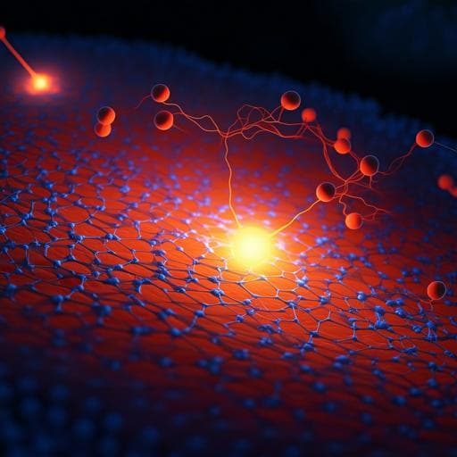
Mixed-state electron ptychography enables sub-angstrom resolution imaging with picometer precision at low dose
Z. Chen, M. Odstrcil, et al.
Discover how researchers Zhen Chen, Michal Odstrcil, Yi Jiang, Yimo Han, Ming-Hui Chiu, Lain-Jong Li, and David A. Muller have achieved groundbreaking low-dose, sub-angstrom resolution imaging with picometer precision. Their innovative mixed-state electron ptychography approach significantly enhances imaging efficiency while reducing radiation exposure, paving the way for new insights in the realm of complex nanostructured materials.
Related Publications
Explore these studies to deepen your understanding
Adjacent work that informs or extends this paper's methodology and findings.

Chemistry
Imaging 3D chemistry at 1 nm resolution with fused multi-modal electron tomography
J. Schwartz, Z. W. Di, et al.

Physics
Low-dose real-time X-ray imaging with nontoxic double perovskite scintillators
W. Zhu, W. Ma, et al.

Medicine and Health
Rapid 3D imaging at cellular resolution for digital cytopathology with a multi-camera array scanner (MCAS)
K. Kim, A. Chaware, et al.
Biology
In vivo volumetric imaging of calcium and glutamate activity at synapses with high spatiotemporal resolution
W. Chen, R. G. Natan, et al.


