
Medicine and HealthNature Communications
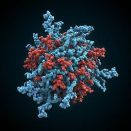
Mechanism of action for small-molecule inhibitors of triacylglycerol synthesis
X. Sui, K. Wang, et al.
Explore the groundbreaking findings on DGAT1, an enzyme crucial for triacylglycerol synthesis, and discover how inhibitors T863 and DGAT1IN1 operate in distinct ways. This study, conducted by Xuewu Sui and colleagues, sheds light on the selective inhibition mechanisms relevant to metabolic diseases and future therapeutic developments.
Related Publications
Explore these studies to deepen your understanding
Adjacent work that informs or extends this paper's methodology and findings.
Medicine and Health
Targeting the coronavirus SARS-CoV-2: computational insights into the mechanism of action of the protease inhibitors lopinavir, ritonavir and nelfinavir
G. Bolcato, M. Bissaro, et al.
Biology
Mechanism of fertilization-induced auxin synthesis in the endosperm for seed and fruit development
L. Guo, X. Luo, et al.

Medicine and Health
An aptamer-based depot system for sustained release of small molecule therapeutics
D. Wang, Y. Li, et al.

Chemistry
Discovery of *Trypanosoma brucei* inhibitors enabled by a unified synthesis of diverse sulfonyl fluorides
B. S. Mantilla, J. S. White, et al.


