
ChemistryNature Communications
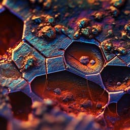
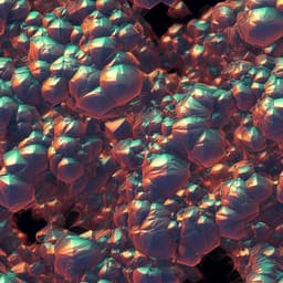
Mechanical cleaning of graphene using in situ electron microscopy
P. Schweizer, C. Dolle, et al.
This groundbreaking research by Peter Schweizer and colleagues unveils a novel site-specific mechanical cleaning technique leveraging in situ electron microscopy. This method promises to effectively eliminate surface contamination from 2D membranes like graphene, enhancing experimental accuracy and paving the way for advances in nanocrystalline graphene synthesis.
Related Publications
Explore these studies to deepen your understanding
Adjacent work that informs or extends this paper's methodology and findings.
Engineering and Technology
Observation of stress corrosion cracking using real-time in situ high-speed atomic force microscopy and correlative techniques
S. Moore, R. Burrows, et al.

Physics
Scaling behavior of electron decoherence in a graphene Mach-Zehnder interferometer
M. Jo, J. M. Lee, et al.
Chemistry
Imaging the microstructure of lithium and sodium metal in anode-free solid-state batteries using electron backscatter diffraction
T. Fuchs, T. Ortmann, et al.

Physics
In situ electron paramagnetic resonance spectroscopy using single nanodiamond sensors
Z. Qin, Z. Wang, et al.


