
Chemistry
Mapping protein binding sites by photoreactive fragment pharmacophores
P. Ábrányi-balogh, D. Bajusz, et al.
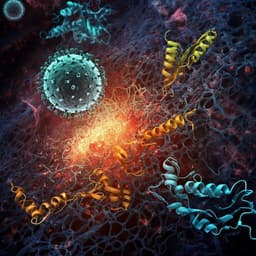
This exciting research, conducted by a team including Péter Ábrányi-Balogh and Zoltán Orgován, presents a novel screening approach that integrates evolutionary optimized fragment pharmacophores with a photoaffinity handle, yielding high hit rates through LC-MS detection. Enhanced by a target-conjugated photocatalyst, this study reveals many fragment hits across six protein targets, showcasing impressive performance over traditional drug discovery methods.
~3 min • Beginner • English
Introduction
Fragment-based drug discovery (FBDD) samples chemical space efficiently with small, polar fragments but detecting their weak, non-covalent binding and mapping the corresponding protein binding sites requires sensitive, often resource-intensive techniques. Photoaffinity labeling has emerged to capture transient interactions, with prior platforms such as fully functionalized fragments (FFFs) and GSK’s PhABits enabling MS-based detection across proteins. Building on these, the authors aim to maximize coverage of experimentally validated fragment-binding pharmacophores using their SpotXplorer design, and to pair such optimized fragments with a diazirine photo-crosslinker to directly map binding sites by intact-protein MS. The study’s purpose is to develop and validate a pharmacophore-optimized photoaffinity fragment (PhP) library, benchmark its performance on tractable and challenging targets (BRD4-BD1, KRasG12D, STAT5B NTD), and improve detection sensitivity via a protein-conjugated photocatalyst, thereby providing experimentally grounded binding-site maps and viable starting points for drug discovery.
Literature Review
The paper situates its work in three strands of literature: (1) FBDD, which achieves higher hit rates by screening small, less complex fragments but faces challenges in sensitivity and structural site assignment; (2) photoaffinity labeling, where FFFs from the Cravatt group and PhABits from GSK demonstrated proteome-wide and targeted fragment capture with MS readouts; and (3) pharmacophore-driven fragment library design, notably the SpotXplorer approach identifying 425 unique binding pharmacophores distilled from 3343 protein–fragment complexes and emphasizing coverage of 2- and 3-point pharmacophore patterns. Prior analyses showed that many fragments act as privileged chemotypes, binding multiple targets while still forming specific interactions in X-ray structures; a Novartis meta-analysis found 63% of fragments never hit, with a minority being multi-target binders. Commercial FFF libraries can be biased toward a few privileged pharmacophores, whereas SpotXplorer libraries distribute coverage more evenly. For KRas, recent work identified fragments and ligands at multiple pockets (Switch II, SOS interface, Switch I/II), yet other surfaces (RBD, dimerization interfaces) remained largely unliganded. For STAT N-terminal domains, emerging structural and virtual screening studies on STAT3 provided a basis to explore the unliganded STAT5B NTD.
Methodology
Library design and synthesis: Fragments were selected from the Enamine primary amine collection, filtered to 10–16 heavy atoms, exactly one primary amine (used solely as the attachment site), and no PAINS. Using SpotXplorer-derived 2- and 3-point pharmacophore models (117 total), fragments were annotated as pharmacophore fingerprints and a diversity picker selected 160 candidates maximizing pharmacophore coverage and diversity. Fragments were coupled to 3-(3-methyl-3H-diazirin-3-yl)propanoic acid via HATU/DIPEA-mediated amide formation (plate-based parallel synthesis). Purification used SPE or prep-HPLC; LC-MS and NMR confirmed products. Synthesis success yielded 100 diazirine-tagged fragments (62% success), covering 88% of the 2-point and 75% of the 3-point pharmacophores.
Screening workflow: One fragment per well was incubated with target protein (15 µL) for 15 min, irradiated for 10 min at 302 or 365 nm, and analyzed by intact-protein MS to detect mass additions indicative of labeling. Hits were categorized by labeling efficiency: strong (>5%), weak (1–5%), multiple binders, oxidized/side reactions, or non-binders (<1%). Benchmarks (carbonic anhydrase II, lysozyme, myoglobin) and three therapeutic targets (BRD4-BD1, KRasG12D, STAT5B-NTD) were screened. BRD4-BD1 and KRasG12D were analyzed on LC-MS-TOF; STAT5B-NTD on a UHPLC-QToF platform to increase accessibility.
Binding site identification and structural methods: Hit labeling sites were mapped by LC-MS/MS peptide mapping (Trypsin/LysC and/or ProAlanase digests). For KRasG12D, HSQC NMR (15N-SOFAST-HMQC) provided residue-level perturbations to guide site assignment. Induced Fit Docking (Schrödinger) modeled binding modes constrained by MS and NMR data. For BRD4-BD1, X-ray crystallography of PhP053 confirmed binding in the AcK site (PDB 8Q34). For STAT5B-NTD, the structure was homology-modeled based on STAT3-NTD.
Photocatalyst enhancement: To boost diazirine labeling efficiencies, an iridium-based photocatalyst (Ir-G2-PEG3-COOH) was conjugated to STAT5B-NTD in aqueous buffer using BOP activation (effective compared to other agents). Upon blue-light irradiation, Dexter energy transfer from the catalyst enhanced crosslinking yields of bound PhP fragments, increasing detection sensitivity and primary hit counts on standard LC-MS platforms.
Functional and cellular assays: A KRas–SOS nucleotide exchange assay with MANT-GDP assessed functional modulation by fragments before and after UV irradiation. Cell viability assays (MTT) tested PhP072 in KRas-dependent PANC-1, SW1990, SW48-PAR, SW48-G12D lines. For STAT5B-NTD, microscale thermophoresis (MST) determined direct binding affinities (Ka), and CellTiter-Blue viability assays evaluated effects in MV4-11 and MOLM-13 leukemia cells.
Analytical conditions: Detailed LC-MS/HPLC gradients, MS acquisition parameters, NMR conditions, crystallization, and data analysis software are reported (including Waters and Agilent platforms; Schrödinger suite for modeling).
Key Findings
- Library performance and coverage: 100 diazirine-tagged fragments (62% synthesis success) covered 88% of 2-point and 75% of 3-point experimentally validated pharmacophores. Benchmark labeling counts: carbonic anhydrase (17 fragments), lysozyme (16), myoglobin (23), with efficiencies comparable or superior to larger prior libraries (e.g., PhABits).
- Hit rates on therapeutic targets: BRD4-BD1 yielded 30 labeling events (>1% across the library; 5 fragment hits confirmed >1% for BRD4-BD1), STAT5B-NTD 26, KRasG12D 25. A subset of fragments showed privileged, selective labeling patterns per protein (e.g., PhP053 favored BRD4-BD1; PhP048 and PhP012 for KRasG12D at 29.4% and 15.8% labeling, respectively; STAT5B-NTD selectively labeled by PhP040, PhP077, PhP065, PhP097).
- BRD4-BD1: Peptide mapping and crystallography (PDB 8Q34) confirmed PhP053 binding in the primary acetyl-lysine site; modeling indicated additional, previously unreported surface-exposed sites (e.g., residues 156–163) for PhP053 and PhP072.
- KRasG12D: Intact MS identified 11 crosslinking compounds; three confirmed hits mapped by LC-MS/MS and HSQC NMR to multiple surfaces, including Switch II pocket (H95/Y96), SOS interface (D33), Switch I (D33), RBD interface (D33), and dimer interface (C118). PhP072 binds distinct sites in the Switch II pocket (H-bonds with D69/E63) and SOS interface (S39/T35/E37). PhP060 targets RBD/dimerization via H-bonds to D33/H27/T35 and π–π to H27; PhP071 binds the dimerization interface (H-bonds R123/T127).
- KRas function and cellular effects: In the KRas–SOS exchange assay, PhP072 minimally affected exchange pre-irradiation but fully restored function to control upon UV-induced crosslinking, consistent with covalent capture at functional interfaces. PhP072 showed antiproliferative activity in KRas-dependent cells with IC50: 17.2 µM (SW1990), 22.6 µM (PANC-1); also tested in SW48-PAR and SW48-G12D.
- STAT5B-NTD: 24 hits identified; two top hits (PhP065, PhP097) labeled >5% without side reactions and bound STAT5B-NTD by MST with Ka = 2.58 µM (PhP065) and 18.06 µM (PhP097). Predicted binding near dimerization interfaces: PhP065 at the handshake dimer cavity (H-bonds Q36/K70), PhP097 near the Ni2+-mediated tetramer interface (H-bond T58, hydrophobic contacts). Cellular IC50 (MV4-11/MOLM-13): PhP065 76.15/100.1 µM; PhP097 56.62/68.22 µM.
- Photocatalyst enhancement: Conjugated Ir-G2-PEG3-COOH increased labeling efficiency and primary hit counts against STAT5B-NTD. Hit classification counts shifted from (no photocatalysis) ≤1%:74, 1–5%:22, ≥5%:2, oxidation:2 to (photocatalysed) ≤1%:40, 1–5%:36, ≥5%:17, oxidation/side reactions:7.
- Comparative efficiency: Versus PhABits, PhP achieved higher hit rates and broader site coverage: for BRD4-BD1, similar pharmacophore coverage with fewer compounds (100 vs 556) and higher hit rate (approx. 5% vs 1.6%); for KRasG12D, PhP hits labeled five distinct sites (vs one with PhABits) with ~7× higher hit rate (5% vs 0.7%).
Discussion
The pharmacophore-optimized photoaffinity fragment (PhP) approach integrates balanced pharmacophore coverage with direct MS-detectable photo-crosslinking to efficiently discover and map fragment binding sites. On a tractable target (BRD4-BD1), PhP confirmed primary-site binders and revealed additional surface-exposed sites that may enable allosteric or orthosteric–allosteric strategies. On a challenging target (KRasG12D), PhP identified fragments at multiple functional and protein–protein interaction interfaces, including Switch I/II, SOS, RBD, and dimerization surfaces—some previously underexploited—demonstrating the method’s ability to experimentally chart allosteric landscapes. For the unliganded STAT5B-NTD, the platform produced the first fragment evidence for targeting dimerization/tetramerization interfaces, corroborated by direct binding (MST) and cellular activity, highlighting tractability of this domain.
The method advances prior platforms by optimizing pharmacophore space coverage (SpotXplorer principles) while retaining the experimental site mapping of photoaffinity screens (FFF/PhABits). Compared with PhABits, PhP delivered higher hit rates and more diverse site discovery with a smaller library. Methodological improvements—compatibility with widely available LC-MS systems and a bioconjugated iridium photocatalyst—enhanced sensitivity, particularly for low-affinity fragment detection. While fragments exhibited on-target binding without irradiation in orthogonal assays (X-ray, NMR, MST), their modest potencies and known promiscuity of diazirine-tagged fragments in cells necessitate cautious interpretation of cellular phenotypes and further target deconvolution.
Overall, PhP efficiently addresses the core FBDD challenge of identifying and mapping viable binding sites early, guiding hit-to-lead efforts with experimentally supported site annotations on both tractable and high-value, historically difficult targets.
Conclusion
This work introduces a 100-member pharmacophore-optimized photoaffinity fragment library that combines broad, balanced pharmacophore coverage with MS-detectable site mapping. The platform effectively identified binders and mapped primary and alternative binding sites for BRD4-BD1, discovered fragments occupying multiple KRasG12D surfaces (including functional and PPI interfaces) with functional assay support and cellular effects, and delivered the first fragment hits against the STAT5B N-terminal domain near its dimerization interfaces, with direct binding and cellular activity. Compared to existing photoaffinity fragment platforms, PhP achieved higher hit rates and explored a greater diversity of binding sites with fewer compounds. Methodological advances—use on standard LC-MS systems and photocatalyst-enabled boosting of diazirine labeling—further democratize and sensitize the approach.
Future directions include: optimizing fragment chemotypes to improve affinity and specificity while minimizing side reactions; refining photocatalyst conjugation and irradiation conditions to balance sensitivity with chemical stability; expanding to broader proteomes/targets; and comprehensive chemoproteomics and genetic perturbation to attribute cellular effects definitively to on-target engagement.
Limitations
- Diazirine labeling efficiencies are typically low (~≤5%), limiting detection sensitivity without enhancement; although photocatalysis improves yields, it increases side reactions (oxidation, water elimination) for certain moieties.
- Potential nonspecific crosslinking/lipophilic interactions and long-lived carbene formation can confound interpretation of site selectivity from intact MS alone.
- Cellular activities of diazirine-tagged fragments may reflect promiscuous engagement; large-scale chemoproteomics suggests such fragments can label many proteins in cells, necessitating careful on-target validation.
- Photocatalyst conjugation was target- and reagent-dependent (BOP effective; others not), and while successful on STAT5B-NTD, generalizability across diverse proteins requires further validation.
- The approach requires UV/visible light irradiation, which may induce protein oxidation/degradation and complicate some assays.
- Some structural assignments relied on docking guided by MS/NMR rather than complete high-resolution co-crystal structures for all hits.
Related Publications
Explore these studies to deepen your understanding of the subject.